Home | About | Contact


Antithrombogenic electrospun scaffolds


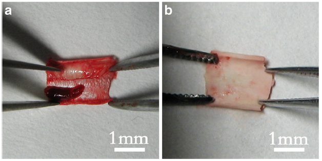
Thrombosis on vascular grafts. (a) Obvious thrombus formation on the vascular graft with smooth topography. (b) Less thrombus formation on the grafts with aligned topography. [Liu et al. BMC Cardiovascular Disorders 2013; 13: 79. This work is licensed under a Creative Commons Attribution 2.0 Unported License].

In applications such as vascular graft, it is vital that the blood flow is not obstructed due to thrombogenic reaction within its lumen. Electrospun scaffold has been found to show low level of adhesion and aggregation of platelets compared to smooth surface or solvent cast scaffold [Ahmed et al 2014]. Milleret et al (2012) found that scaffold composed of fibers less than 1 µm triggered very low coagulation with negligible platelet adhesion. However, larger fiber diameter (2- 3 µm and 5 µm) triggered higher thrombin formation and platelet adhesion. Liu et al (2013) compared the hemocompatibility of electrospun polyurethane and smooth surface vascular grafts in a rat abdominal aorta model. After fifteen minutes of implantation, there was less thombus formation in the electrospun scaffold compared to the smooth surface graft. In a 7 week implantation of electrospun poly(L-lactic acid)-co-poly(ε-caprolactone) scaffold into a rabbit inferior superficial epigastric vein model, no blood coagulation occurred in the lumen of the scaffolds although no endothelial cells were also found [He et al 2009]. However, for longer duration in vivo study, implanted electrospun poly(L-lactic acid) (PLLA) acellular scaffold in a rat common carotid artery showed significant intimal thickening in the lumen after 60 days [Hashi et al 2007]. For short term implant or contact with blood, electrospun fibers with diameter less than 1 µm was able to retard thrombus formation. For longer term, other strategy is needed to maintain patency of the graft.

Property of the electrospun material may be modified through the use of additives to increase its anti-thrombogenicity. Electrospun silk fibrous scaffold demonstrate low thrombogenic potential that is similar to PTFE in vitro. Thrombogenic potential of electrospun silk fibrous scaffold can be further lowered by the conjugation with heparin [Seib et al 2014]. Dhandayuthapani et al (2012) found that increasing the concentration of single wall carbon nanotubes (SWCNTs) in electrospun zein was able to decrease platelet adhesion. Other anti-thrombogenic biomolecules or drugs may also be added to the electrospun scaffold. Del Gaudio et al (2013) were able to reduce platelet adhesion on electrospun polycaprolactone (PCL) scaffold in a 6 hour period by loading aspirin into the fibers although their diameter were more than 3 µm. Hong et al (2009) reported just 40% patency for poly(ester urethane)urea (PEUU) electrospun grafts and 67% patency for non-thrombogenic poly(2-methacryloyloxyethyl phosphorylcholine-co-methacryloyloxyethyl butylurethane) (PMBU)/PEUU blended electrospun grafts after 8 weeks implant in a rat infrarenal aorta model. The relatively better performing PMBU/PEUU blend showed continuous endothelium in the lumen with no thrombogenic deposition and intimal hyperplasia but electrospun PEUU grafts showed occluded lumen. To further reduce thrombogenesis, non-thrombogenic phospholipid copolymer has been grafted to the surface of electrospun PEUU instead of blending. This resulted in a much better performance of 92% patency at 8 weeks in the same animal model with the same continuous endothelium lining observed. Hashi et al (2010) bonded Hirudin to electrospun PLLA fibers with the help of di-amino polyethylene glycol (PEG). Despite having a fiber diameter of about 2 µm, the presence of PEG and Hirudin-PEG added to PLLA was able to inhibit platelet adhesion on the fiber surface as compared to pure PLLA. In vivo study showed that the Hirudin-PEG grafted PLLA was able to maintain patency at 6 months. Their study also showed endothelialisation on the luminal surface with complete endothelial coverage at 1 month and 6 months. Wang et al (2020) constructed an anti-thrombogenic liquid gating membrane-based catheter (LGMC) with electrospun tube as the main structural support. This catheter uses a porous membrane with a liquid filling up the pores in the membrane (gating liquid) to create a continuous wall. It is important that the affinity between the gating liquid and the porous membrane is strong so that the liquid does not leak out from the pores. Increasing the thickness of the tube wall by having a longer electrospinning duration increases the pressure threshold. However, increasing the solution concentration which increases the fiber diameter decreases the pressure thresholds exponentially. This may be due to larger pore sizes of larger diameter fibers. Wang et al (2020) showed that electrospun polyvinylidene fluoride (PVDF) porous tube with perfluorodecalin, Krytox 100, Krytox 103, and silicone oil 500 as gating liquid were able to resist coagulation and clot formation better than bare PVDF tube. The gating liquid may also be used as a carrier for drugs to be released at the surgical site.

In native blood vessel, a layer endothelial cells lines the wall of the lumen and this is thought to prevent thrombus formation. In vitro studies have shown that a layer of mesenchymal stem cells (MSC) or endothelial cells (ECs) were able to lower platelet adhesion/aggregation while smooth muscle cells (SMCs) performed poorly [Hashi et al 2007]. Similarly, having a layer of MSCs or ECs in the lumen of graft may be the solution for long term patency of the graft. Hashi et al (2007) seeded mesenchymal stem cells (MSC) onto a sheet of electrospun poly(L-lactic acid) (PLLA) scaffold and physically roll into a tube before implantation into a rat common carotid artery. After 60 days, the MSC-seeded vascular grafts showed little intimal thickening in contrast to intimal thickening in the lumen of acellular grafts. Niu et al (2021) constructed a bilayered electrospun tubular vascular scaffold with the inner layer made of electrospun hyaluronic acid (HA) fibers and the outer layer made of electrospun collagen fibers. The bilayered vascular scaffold was cross-linked with glutaraldehyde vapor. To mimic native vascular graft, the inner lumen of the scaffold was seeded with endothelial cells (EC) and the outer surface was seeded with smooth muscle cells (SMC). Platelet adhesion test by exposing the scaffolds to arterial flow showed that bare scaffolds without endothelial cells coverage resulted in numerous platelets adhesion. In contrast, scaffolds with endothelial cells coverage in the lumen had significantly less platelets adhesion. In vivo tests on a rabbit carotid artery model showed that the bilayered scaffold with EC and SMC seeded was able to maintain a mechanical strength close to that of native artery after 6 weeks. In the same period, all cellularized bilayered scaffolds remained patent and their lumen diameter unchanged. Therefore, this cellularized tubular HA/collagen nanofibrous scaffold has the potential to be used as a vascular graft.


Published date: 24 November 2015
Last updated: 15 November 2022

Comments
[+]


Google Ad.