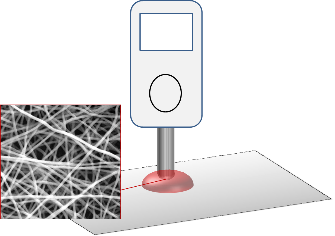
A biosensor is an analytical device which uses a biological element to recognize a target in the environment and give out a signal that can be processed into a usable reading. High surface area of electrospun fibers makes it useful in exposing biological elements to the environment for more sensitive detection. A wide array of encapsulation techniques are available for loading biological elements onto electrospun fibers, each with its advantages and disadvantages. Other than as carrier for biological elements, electrospun fibers may also be used as a component to improve the performance of biosensors.
Carrier for biological elements
There are several ways in which electrospun fibers may be used as a carrier for biomolecules. Direct blending where the biomolecules are added to the solution for electrospinning is commonly used as this is a very straight forward process. For more secure attachment of biomolecules to the electrospun fiber matrix, chemical methods may be employed such as covalent bonding or hydrogen bonding.
Blending has been shown to be an effective method of using electrospun fibers as carriers for functional biomolecules. Guo et al (2011) used collagen as carrier for hemoglobin (Hb) for catalytic reduction of hydrogen peroxide (H2O2). Electrospun fibers with Hb were coated on bar glassy carbon electrode. Their study showed that there is efficient transfer of electron from encapsulated Hb to the electrodes. However, the electron transfer is affected by the thickness of the electrospun membrane and the extent of cross-linking. Since collagen is non-conducting, beyond an optimum thickness, the speed of electron transfer will be reduced. As for cross-linking, too extensive and it will denature the proteins and affect the electron transfer. The resultant amperometric response of this biosensor to H2O2 concentration exhibits a linear output from ranging from 5 x 10⁻⁶ mol L-1 to
30 x10-6 mol L-1, with a detection limit of 0.37 x 10⁻⁶ mol L-1. While blending is an easy method for introducing functional biomolecules into a nanofibrous structure, it has an inherent risk of unwanted leaching during usage. Therefore, stronger bonding between the biomolecules and the nanofiber is sometimes preferred.
Electrospun fibers with functional groups can be used for immobilising biomolecules. Otherwise, there are alternative methods for introducing functional groups such as plasma treatment and UV irradiation. Saha (2015) constructed an electrospun-based membrane for the detection of E. coli K-12. For their biosensor, electrospinning was carried out to product nylon 6 nanofibrous membrane. Surface polymerization was used to coat the nylon 6 membrane with conductive polyaniline (PANi). Antibody goat polyclonal to E. coli was immobilized onto the conductive nylon 6/PANi membrane for detection of E. coli K-12. The resultant biosensor has a linear detection range for E. coli K-12 from 101 to 105 CFU. The time taken for detection of E. coli is very fast at 20 mins compared to other conventional biosensors which typically takes more than 2 hours. Mondal et al (2014) used oxygen plasma treatment to introduce functional groups on TiO2 nanofibers derived from sintering electrospun TiO2 precursors and polyvinyl pyrrolidone (PVP). The functional group permits covalent bonding of cholesterol esterase (ChEt)
and cholesterol oxidase via N-ethyl-N0-
(3-dimethylaminopropyl carbodiimide) and N-hydroxysuccinimide (EDC-NHS) chemistry. The resultant biosensor was able to detect esterified cholesterol with a detection limit of 0.49 mM, excellent sensitivity (181.6 µA/mg dL-1/cm2) and rapid detection (20 s). Hoy et al (2019) used spot oxygen plasma treatment on electrospun polystyrene (ESPS) microfibers membrane to introduce specific hydrophilic zones. Antigens are immobilised on these hydrophilic zones for detection of antibodies specifically against Middle East respiratory syndrome coronavirus (MERS-CoV). Fluid flow through these hydrophilic zones is expected to be faster against the broader hydrophobic surfaces. Embedded within a housing, the plasma treated membrane is first immobilized with the antigen, blocking agent is then held for a minute and passed through before the antibody is captured for identification. The whole process of preparation and detection took less than 6 min. The limits of detection (LOD) of this membrane for MERS-CoV is about 200µg/mL.
Conductivity enhancement
Uniaxial nature of fibers may be used to improve signal conduction produced by the biological element. Li et al (2014) used a mixture of electrospun-derived carbon nanofiber, Nafion and laccase for the detection of catechol, a organic compounds used in the production of pesticides, flavors and fragrances. The constructed biosensor demonstrated a linear range of 1 to 1310 µM, a sensitivity of 41 µA.mM-1 and detection limit as low as 0.63 µM. This performance compares well to other reported laccase-based biosensors. The better performance may be attributed to the presence of electrospun-derived carbon nanofibers which enhanced the conductivity of the composite and hence a faster electron transfer. In the construction of the composite, the electrospun-derived carbon nanofibers underwent ultrasonication to break it into short strands. The short strands of carbon nanofibers were mixed with Nafion and laccase before loading onto glass carbon electrode. The biosensor showed excellent selectivity towards catechol with no response to other tested phenolic compounds such as epicatechin, gallic acid, guaiacol, phenol and aminophenol. Stability of the biosensor over 30 days were good in 0.2 M air-saturated acetate buffer (pH 4.0) at 4 °C with satisfactory recovery in real water samples and good repeatability.
Release agent
Enzyme, proteins and many biomolecules are susceptible to degradation under environmental condition and may lose its activity during storage. For some biosensor device, the biomolecules are only needed when the feed solution is present. Electrospun nanofibers are a good carrier for quick release of enzyme for applications such as microfluidic detection chip. High surface area of the nanofibers allows rapid dissolution in appropriate water or solvents. Dai et al (2012) used water soluble electrospun polyvinyl pyrrolidone nanofibers as carrier for horseradish peroxidises (HRP). Placed in a microfluidic chip, PVP nanofibers loaded with HRP readily dissolve to release HRP when the aqueous sample passed through it. The activity of HRP was found to be reduced by 20% immediately after electrospinning. After storage over 280 days, 40% of its activity remained.
Published date: 05 September 2017
Last updated: 21 April 2020
▼ Reference
-
Dai M, Jin S, Nugen S R. Water-Soluble Electrospun Nanofibers as a Method for On-Chip Reagent Storage. Biosensors 2012; 2: 388. Open Access
Open Access
-
Guo F, Xu X X, Sun Z Z, Zhang J X, Meng Z X, Zheng W, Zhou H M, Wang B L, Zheng Y F. A novel amperometric hydrogen peroxide biosensor based on electrospun Hb-collagen composite. Colloids and Surfaces B Biointerfaces 2011; 86: 140.
-
Hoy C F O, Kushiro K, Yamaoka Y, Ryo A, Takai M. Rapid multiplex microfiber-based immunoassay for anti-MERS-CoV antibody detection. Sensing and Bio-Sensing Research 2019; 26: 100304.
Open Access
-
Li D, Pang Z, Chen X, Luo L, Cai Y, Wei Q. A catechol biosensor based on electrospun carbon nanofibers. Beilstein J. Nanotechnol. 2014; 5: 346.
Open Access
-
Mondal K, Md. Ali A, Agrawal V V, Malhotra B D, Sharma A. Highly Sensitive Biofunctionalized Mesoporous Electrospun TiO2 Nanofiber Based Interface for Biosensing. ACS Appl. Mater. Interfaces 2014; 6: 2516.
-
Saha T. Detection of Foodborne Biohazards Using Antibody Modified Electrospun Conducting Fibres. Master of Sciences in Food Science Thesis. The University of Guelph. Canada. 2015.
▲ Close list
 ElectrospinTech
