Influence of fiber alignment on cells and tissue regeneration
The resemblance of electrospun nanofibers scaffold to natural extracellular matrix (ECM) has made it a widely used tool in the study of cell behaviour. Since many extracellular matrixes (ECM) are made out of organized collagen fibers, this has led to many studies on the effect of aligned electrospun fibers on various cell types. Several studies have shown that cells cultured on aligned electrospun fibrous substrate, exhibit contact guidance. This is particularly useful in neural engineering where the suitability of the scaffold is determined in part from the length neurite extension. In highly aligned nanofiber substrate, neurite outgrowth can be as much as 20% longer than that on random fibers [Corey et al 2007]. Schwann cells were also shown to elongate along the direction of fiber orientation [Corey et al 2007, Gnavi et al 2015]. When compared to film matrix of the same material composition, aligned nanofibers comprising of carbon nanotube/polycaprolactone/gelatin showed greater NRG1 and P0 protein expression levels in Schwann cell [Tsai et al 2014]]. However, Gnavi et al (2015) reported lower proliferation of Schwann cells on aligned gelatin fibers compared to randomly oriented gelatin fibers. Fiber orientation also did not affect the neurite length 50B11 sensory neurons-like cells although it does induce neurite orientation [Gnavi et al 2015]. Fee et al (2016) examined the gene expression of NIH3T3 cells fibroblasts on aligned and randomly oriented electrospun polycaprolactone (PCL)/Gelatin nanofibers. In agreement with other studies, the fibroblasts were oriented in the direction of the aligned fibers but showed little orientation preference on randomly oriented fibers. Up-regulated genes for fibroblasts seeded on aligned fibers when compared to random fibers are associated with actin production, actin polymerization and focal adhesion formation. This may be due to the deformation and stretching of the cell nucleus to align with the fiber orientation which necessitates greater actin production and focal adhesion.
Cell alignment due to fiber orientation is also dependent on the fiber diameter and cell type. For adult human dermal fibroblasts, Liu et al (2009) found that a minimum diameter of 0.97 µm is required for cell orientation to occur. At lower fiber diameter, the aspect ratio of the cell is comparable to film. On the contrary, for endothelial cells, aligned fiber diameter over 1.2 µm seems to have less influence on cell orientation. Using electrospun aligned polycaprolactone (PCL)/collagen fibers with different fiber diameters (100 nm, 300 nm and 1200 nm), Whited and Rylander (2014) found that influence on fiber orientation on primary human umbilical vein endothelial cells (HUVEC) orientation was weakest for 1200 nm fibers. There was no significant difference in fiber directed cell orientation for diameter of 100 nm and 300 nm.
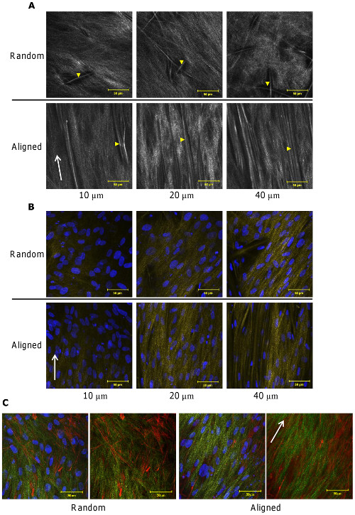
Apart from contact guidance of the cells on aligned fibers, the deposition of collagen fibers were also found to align with the orientation of the fibers. Using second harmonic generation (a minimally invasive, two photon based technique for visualization of collagen) to examine the the deposition of normal human dermal fibroblasts on electrospun polycaprolactone microfibers (about 8 µm), collagen appeared to be more organized and oriented on aligned microfibers while collagen deposited on random microfibers did not show any preferential organization. Such collagen distributions were observed down to 100 µm depth within the scaffolds [Delaine-Smith et al 2014]. In vivo study of implanted aligned fibrous scaffold in a subcutaneous rat model also showed that collagen fiber deposited was aligned whereas collagen fibers deposited on randomly oriented fibrous scaffold was not [Ifkovits et al 2010]. Organization of deposited collagen fibers are likely to be affected by local alignment of the electrospun fibers. Xie et al (2010) showed that the secreted collagen type I showed high degree of organization in the radially aligned membrane in contrast to the haphazard arrangement for random fibers membrane.
Fibroblast-secreted collagen and scaffold fibres were visualised using SHG emissions collected at depths of 10-40 µm (A). Yellow arrow heads identify PCL fibres of the scaffold. Fibroblast-seeded constructs were fixed and visualised via SHG emissions (collagen and scaffold fibres in yellow) and DAPI staining (cell nucleus in blue) at depths of 10-40 µm (B). Fixed constructs were also visualised for fibroblast cytoskeleton (phalloidin-TRITC in red) proximity to secreted collagen (SHG in green) (C). SHG was obtained using 800 nm illumination and all images were collected after 21 days of culture. White arrows indicate scaffold fibre orientation [Delaine-Smith RM, Green NH, Matcher SJ, MacNeil S, Reilly GC (2014) Monitoring Fibrous Scaffold Guidance of Three-Dimensional Collagen Organisation Using Minimally-Invasive Second Harmonic Generation. PLoS ONE 9(2): e89761. doi:10.1371/journal.pone.0089761. This work is licensed under a Creative Commons Attribution 3.0 Unported License].
Zhu et al (2019) investigated the effect of adding kartogenin (KGN) to electrospun aligned polycaprolactone (PCL) for treating rotator cuff tear (RCT). KGN is a small molecule drug that was found to promote chondrocyte differentiation and is blended into PCL solution before electrospinning. Animal studies were carried out on a rat model by severing its rotator cuff tendon and removal of its fibrocartilage. The tendon was repaired to its anatomic footprint by transosseous repair with the aid of KGN loaded electrospun aligned PCL membrane. The collagen fibrils were better organised in the groups with aligned electrospun membranes.
Where the ECM of a tissue is made out of oriented fibers, culturing their cells on aligned nanofibrous scaffold may show responses that mimics that of the cells in its native environment. The periodontal ligament (PDL) is one such tissue which is made out of organized collagen fibers. Shang et al (2010) cultured rat PDL cells on aligned parallel and cross electrospun fibrous poly (lactide-co-glycolide) (PLGA) scaffolds and their metabolic activities are significantly higher than randomly oriented fibrous scaffold and solvent cast film. The cells were also extended and aligned to the aligned fibers with a spindle-like shape in contrast to the polygonal shape of cells cultured on randomly oriented fibrous scaffold. Similar cell shape was also observed in the study by Liu et al (2015) where rabbit anuulus fibrosus stem cells (AFSCs) were cultured on aligned poly(ether carbonate urethane)-urea (PECUU) fibers. Further production of collagen-I and glycosaminoglycans (GAG) was also found to be significantly more in AFSCs cultured on aligned fiber scaffold compared to random oriented fibrous scaffold. The aligned fibrous scaffold was able to mimic the outer anuulus fibrosus which comprised of highly organized, aligned collagen fibers. Cells in the native environment are elongated fibroblast-like and aligned in the direction of collagen fibers.
You et al (2022) investigated the effect of nanofiber orientation on cell glucose metabolism. Using bone marrow mesenchymal progenitor cell line, ST2 cells, cultured on aligned and randomly oriented polycaprolactone (PCL), they found that cells cultured on randomly oriented fibers showed greater glycolytic capacity such as enhanced glucose consumption and lactate production compared to aligned fibers. Similar to other studies, the cells showed preferential alignment in the direction of the aligned fibers with the nucleus of cells being rounded on random fibers and elongated on aligned fibers. The myosin filaments and intracellular tension was found to be greater in cells cultured on random fibers with greater expression of phospho-myosin II (Ser1943). It is inconclusive whether greater glycolytic activity when cells are cultured on randomly oriented fibers is beneficial as it may be due to stress on the cells. Nevertheless, it provide further evidence on nanofiber orientation influencing cellular activity.
Formation of vascular tubules requires the endothelial cells to come together and align in a given direction. Thus, scaffold-directed endothelial cell alignment may be the first step towards encouraging angiogenesis. Studies by Montero et al (2014) and Brown (2012) have shown that aligned fibers were able to guide endothelial cells growth and their formation to functional vessels while random fibers results in scattered endothelial cells. Brown (2012) further studied the effect of aligned fiber diameter on the quantity of in vivo blood vessel formation in rat spinal cord injury. Using polydioxanone aligned fibers of average diameter 1 µm and 2 µm, it was found that the smaller fiber diameter yield more blood vessels. Further comparative studies between aligned and random fibers on endothelial cells behaviour was carried out by Gaharwar et al (2015). Their study uses human umbilical vein endothelial cells cultured on electrospun poly(glycerol sebacate) (PGS)/poly(ε-caprolactone) (PCL) with diameters of 3.4 µm and 4.7 µm for random and aligned fibers respectively. Apart from alignment of cell nuclei and actin fibers on the aligned fibers, cell proliferation and expression of CD31, a major determinant in angiogenesis, was also greater on the aligned fibers compared to the random fibers. This provided greater evidence that aligned fibers is able to encourage angiogenesis. Using near field electrospinning, Xue et al (2014) formed an array of aligned gelatin fibers on PDMS to test its influence on endothelial cells (ECs). Hydrated electrospun gelatin fiber has a height of 1 µm and width of 4 µm. They found that the ECs formed cell strings on the fiber arrays and each cell string resembles pre-capillary cord. However, the cells did not form into lumen within 20 hrs of culturing. Such string like cell organization was not observed on featureless 2D PDMS surface. While it is not clear why such cell organization take place, it could be due to the more favourable attachment surface provided by gelatin instead of PDMS and this encourages the cells to wrap round the fibers. In directing the physical morphology of the cells, the fibers may facilitate vascular formation by endothelial cells.
(a) Assembly of endothelial "cell strings" over a large area of gelatin fibril pattern. (b) An image slice of the reconstructed 3-D conformation of a selected cell string. [Xue et al PLoS ONE 2014; 9: e93590. This work is licensed under a Creative Commons Attribution 4.0 International.]
Fiber alignment has been shown to facilitate stem cell differentiation to particular cell lineage. With differentiation of mesenchymal stem cells (MSC) towards ligament fibroblast-like cells, mechanical stimulation alone is insufficient. Subramony et al (2013) showed that the mechanically stimulated MSC needs to be cultured on scaffold with aligned fibers to induce differentiation while scaffold with unaligned nanofibers does not. Evidence of differentiation of ligament fibroblast-like cells from a combination of mechanical loading and nanofiber alignment came from a measurement of the collagen type I:III ratio which was approximately 8:1 after 28 days. This is ratio is closest to native ligament collagen Type I:III ratio of about 7:1 compared to unloaded scaffolds and scaffold with unaligned fibers. Other expression of fibroblast-related markers by MSCs on aligned and mechanically loaded scaffold includes fibronectin expression and tenascin-C after 14 days [Subramony et al 2013]. Just as aligned fiber scaffold have been shown to facilitate neurite outgrowth in nerve applications, aligned fiber scaffold has also been shown to enhance embryonic stem (ES) cells differentiation into neural lineages. Xie et al (2009) seeded mouse embryonic stem on both aligned polycaprolactone (PCL) fibers and randomly oriented fibers. Cultured in media containing 4-/4+ retinoic acid treatment protocol, cells were induced to form embryoid bodies (EB) before seeding on aligned and random fiber samples. After 14 days, EBs on aligned fibers showed higher Tuj1 (early neurons) and O4 (oligodendrocytes) markers and lower numbers of GFAP (glia fibrillary acidic protein)-positive cells. More astrocytes were also found to be present around the embryoid bodies (EBs) cultured on random fibers compared to aligned fibers. Seonwoo et al (2018) demonstrated the influence of fiber alignment on dental pulp stem cells (DPSCs) morphology and linkages with neighbouring cells. Their study has shown that having 0.1% reduced graphene oxide (RGO) in electrospun polycaprolactone (PCL) significantly improved DLSCs neurogenic differentiation. On random RGO/PCL electrospun fibers, the differentiated neurites exhibited multipolar structures with connection with neighbouring cells. On aligned RGO/PCL electrospun fibers, the differentiated neurites exhibited bipolar structures and connects to other cells in a linear direction. Seonwoo et al (2018) proposed that random fibers is suitable for the regeneration of the central nervous system due to its influence on differentiated neurites morphology and linkage characteristic. For aligned fibers, it is suitable for regeneration of the peripheral nervous system based on differentiated neurites linear alignment.
There are mixed results in the differentiation of stem cells to osteoblastic lineage between cells cultured on aligned and randomly oriented fibers. Ma et al (2011) showed that bone marrow stromal (BMS) cells cultured on aligned poly(L-lactide) (PLLA) nanofibers exhibits significantly greater calcium content at 21 days for cells on aligned PLLA nanofibers compared to randomly oriented nanofibers. However, there were no significant difference in the ALPase activity and expression of osteopontin (OP) and osteocalcin (OC). However, in the study by Doustgani and Pedram (2016) using MSCs cultured on polylactide/poly (vinyl alcohol)/ Calcium carbonate nanoparticles electrospun fibers, ALP activity was the highest for aligned fibers followed by random fibers and lastly, TCPs at 21 days.
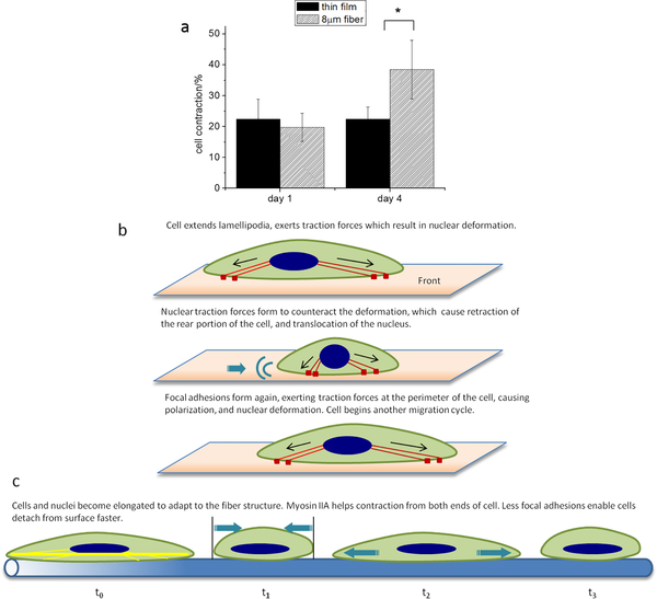
Orientation of the fibers is able to facilitate cell migration. Using a radially aligned nanofiber membrane, Xie et al (2010) showed enhanced migration of dural fibroblast towards the center of the membrane compared to membrane with randomly oriented fibers. Shang et al (2010) showed that rat PDL cells migration on aligned fibers (parallel and cross) was also significantly faster than randomly oriented fibrous scaffold and solvent cast film. Qin et al (2015) reported a detailed study of fibroblast movement on aligned 8 µm diameter polymethylmethacrylate (PMMA) fibers and cast PMMA film. On a fibrillar surface the cells adopted a cytoplasm contraction and expansion mechanism which is similar to that of muscle cells with the nucleus remained highly deformed during migration. On a flat surface, the cells migrate through a sequence of lamellipodia extension, nuclear deformation and retraction of the rear end of the cell. The migration mechanism adopted by the cells on the fibrillar surface allows them to double the contraction amplitude while the contraction of the cells migrating on the flat surface remains unchanged.
Cell contraction associated with migration on thin film and 8 µm fiber-(a) Measurement of cell contraction on micron fibers and flat films at day 1 and day 4. Cell contraction on micron fiber is defined as: (cell length at t0- cell length at t1-)/cell length at t1- *: P<0.001. (b) Illustration of cell contraction when migration on flat film. (c) Illustration of cell contraction when migration on 8 µm fibers at day 4 [Qin et al. PLoS ONE 2015; 10: e0119094].
Cell movement on the fibers are sensitive to any disruption to its path. Johnson et al (2009) showed that with human glioma cell that pauses between cell movement often coincides with fiber-fiber intersection even if the angle of misalignment is small (~20 °). Glioma cell migration speed on aligned fibers was much faster than on random fibers with velocity of 4.2 µm/h compared to 0.8 µm/h.
A study using breast cancer cells showed that influence of fiber alignment on the epithelial - mesenchymal transition (EMT). Saha et al (2012) tested the behavior of nonaggressive luminal cancer cells (MCF-7), aggressive basal cells (MDA-MD-231) and mammary tumor cell (H605) on electrospun aligned and randomly oriented polycaprolactone fibers with diameter of 1.8 µm and 2.0 µm respectively. Aggressive MDA-MD-231 cancer cells were found to align and elongated along the fiber axis but nonaggressive MCF-7 showed random orientation on aligned fibers. Mouse mammary tumor cells (H605) cells cultured on aligned fibers showed significant upregulation in the gene expression of Cytokeratin (Ck14), smooth muscle actin, TGFβ, Snail, fibroblast specific protein and Smad3 while less expression was observed in cells cultured on randomly oriented fibers. TGF? and Snail are well known EMT inducers while Ck14 are known to activate aggressive basal-like breast cancer cells [Saha et al 2012]. Further, Nelson et al (2014) found that on aligned PCL nanofibers (diameter of 795 nm), MDA-MB-231 cells migrated significantly further in the presence of CXCL12 gradient compared to MCF-7 and MCF-10A (normal mammary epithelial) cells. Migration of all cell types on aligned nanofibers is greater than randomly aligned nanofibers. In the progression of breast cancer tumor, the extracellular matrix (ECM) transform from random fiber organization to highly aligned ECM which encourages migration and invasion. Aligned electrospun nanofibers scaffold may be used to mimic such fiber organization to replicate in vivo behavior such as rapid cell migration.
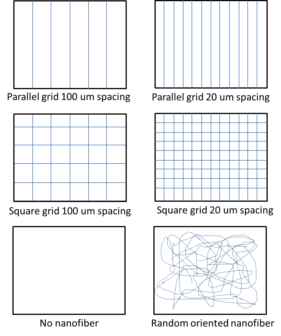
Most studies carried out on electrospun aligned fiber scaffold are on tightly packed aligned fibers. However, the spacing between the aligned fibers may also influence cell behaviour. In this case, there will be an underlying smooth substrate where fibers are spaced out individually. In a study by Fuh et al (2013), the behavior of embryonic kidney cells were tested on fiber (diameter about 700 nm) grids formed on polypyrrole substrates as shown in the figure below. The study showed distinct cell alignment on the parallel grids with fiber-fiber spacing of 20 µm demonstrating better cell orientations than 100 µm spacing. No cell orientation was recorded for other substrates. Investigation of cell spreading showed that the spreading is the same for parallel grid and square grid of the same spacing. However, cell spreading is reduced when the spacing between the fibers are reduced for both square and parallel grids. In separate study by Park et al (2011) using dermal fibroblasts on randomly oriented and aligned polycaprolactone (PCL) fibers, they found that cell alignment is better with higher density of aligned fibers below the cells. Their study showed that cell guidance is more effective when the fiber spacing is similar or smaller than the cell size.
Fiber grid patterns. Fiber grid with 20 µm spacing shows better influence on cells than 100 µm spacing [Fuh et al 2013].
Most investigations on the effect of aligned on cell orientation are based on flat, planar surface. For tissues such as muscles, the ECM is in a 3D form. Therefore, a 3D scaffold comprising of aligned fibers with adequate inter-fiber spacings will be a closer match to muscle ECM. However, aligned fibers will not be able to maintain an open structure without any support. Zhou et al (2018) used a trough collector comprising of a flat base and slanted walls to gather electrospun fibers along the depth of the collector. The collected fibers were aligned wall to wall to form a 3D structure supported by the walls. Fibroblasts cultured on this supported structure were found to be distributed throughout the fibrous matrix and aligned according to the fiber orientation. In this case, the supporting structure purpose is to hold the electrospun fibers in a 3D spatial form only while allowing the cultured cells to populate the scaffold.
Cell behaviour is often influenced by various factors such as surface topography, mechanical stress or fluid shear flow. While cell behaviour may show favourable response when tested on a single factor, the optimum substrate may be different when various factors are combined. Veleva et al (2009) found that proliferation of human blood outgrowth endothelial cells (HBOEC) and human umbilical vein endothelial cells (HUVEC) on electrospun terpolymer (made of hexylmethacrylate (HMA), methylmethacrylate (MMA), and methacrylic acid (MAA)) aligned fibers (majority diameter in the range 4 to 8 µm) were lower than randomly aligned fibers (majority diameter in the range 4 to 12 µm) although both aligned and randomly oriented fibrous scaffold performed better than cast films. While this study may suggest that randomly aligned fibers are better for endothelial cells, another study which includes fluid flow showed that aligned fibers may be better. Whited and Rylander (2014) cultured primary human umbilical vein endothelial cells (HUVEC) on electrospun PCL/collagen scaffolds subjected to fluid flow. Comparing quantity of cells which remained attached on electrospun scaffold with randomly oriented fibers and aligned fibers after 60 minutes of shear stress (20 dyne/cm2), 95% of cells remained adhered to the aligned fibrous scaffold while only 60% of cells remained on randomly oriented fibrous scaffold. In their studies, the diameter of the fibers (100 nm and 300 nm) did not have a significant impact on cell adhesion and retention. Interestingly, their study showed that fiber guided cell orientation was only evident when the fiber diameter is 100 nm and 300 nm and not for 1200 nm diameter fiber.
Cells cultured on aligned fibers have been shown to exhibit varying degrees of orientation along the length of the underlying fibers. With cell electrospinning, it has been shown that it is possible to physically orientate the cells along the length of the fiber. Yeo et al (2023) prepared a cell-laden bioink containing alginate and polyethylene oxide (PEO) (A2P3) in water. Mesenchymal stem cells (MSCs) and smooth muscle cells (SMCs) were added to the A2P3 aqueous solution prior to electrospinning and 3D cell printing (CP). Cell viability after 7 days of culture was found to be more than 90% for both CP scaffold and electrospun scaffold. To produce aligned cell-containing fibers, a parallel electrode collector setup was used. An advantage of this setup over a rotating drum collector setup is that this method would not introduce additional stretching force on the electrospinning jet which may cause additional stress to the embedded cells. The cell-printed scaffold had a plain bulk structure and the cells had a round shape while cells in the electrospun scaffold showed elongation morphology along the length of the fiber. Cells on electrospun scaffold showed higher F-actin aspect ratios and orientation factor and the SMCs showed greater expressions of fibronectin, collagen I and collagen (IV). Therefore the ability of electrospinning to produce finer structure compared to cell printing was able to produce a scaffold more conducive for cell expression.
Published date: 10 November 2015
Last updated: 28 May 2024
Brown D E. Angiogenesis in Response to Varying Fiber Size in an Electrospun Scaffold In Vivo. MSc Thesis. Virginia Commonwealth University 2012.
Open Access
Corey J M, Lin D Y, Mycek K B, Chen Q, Samuel S, Feldman E L, Martin D C. Aligned electrospun nanofibers specify the direction of dorsal root ganglia neurite growth. J Biomed Mater Res 2007; 83A: 636.
Delaine-Smith R M, Green N H, Matcher S J, MacNeil S, Reilly G C. Monitoring Fibrous Scaffold Guidance of Three-Dimensional Collagen Organisation Using Minimally-Invasive Second Harmonic Generation. PLoS ONE 2014; 9(2): e89761. doi:10.1371/journal.pone.0089761
Open Access
Doustgani A, Pedram M S. Preparation and investigation of polylactic acid, calcium carbonate and polyvinylalcohol nanofibrous scaffolds for osteogenic differentiation of mesenchymal stem cells. Nanomed. J. 2016; 3: 109.
Open Access
Fee T, Surianarayanan S, Downs C, Zhou Y, Berry J. Nanofiber Alignment Regulates NIH3T3 Cell Orientation and Cytoskeletal Gene Expression on Electrospun PCL+Gelatin Nanofibers. PLoS ONE 2016; 11(5): e0154806. doi:10.1371/journal.pone.0154806.
Open Access
Fuh Y K, Chen S Z, He Z Y. Direct-write, highly aligned chitosan-poly(ethylene oxide) nanofiber patterns for cell morphology and spreading control. Nanoscale Research Letters 2013; 8: 97.
Open Access
Gaharwar A K, Nikkhah M, Sant S, Khademhosseini A. Anisotropic poly (glycerol sebacate)-poly (ε-caprolactone) electrospun fibers promote endothelial cell guidance. Biofabrication 2015; 7: 015001.
Open Access
Gnavi S, Fornasari B E, Tonda-Turo C, Laurano R, Zanetti M, Ciardelli G, Geuna S. The Effect of Electrospun Gelatin Fibers Alignment on Schwann Cell and Axon Behavior and Organization in the Perspective of Artificial Nerve Design. Int. J. Mol. Sci. 2015; 16: 12925.
Open Access
Ifkovits J L, Wu K, Mauck R L, Burdick J A. The Influence of Fibrous Elastomer Structure and Porosity on Matrix Organization. PLoS ONE 2010; 5(12): e15717. doi:10.1371/journal.pone.0015717.
Open Access
Johnson J, Nowicki M O, Lee C H, Chiocca E A, Viapiano M S, Lawler S E, Lannutti J J. Quantitative Analysis of Complex Glioma Cell Migration on Electrospun Polycaprolactone Using Time-Lapse Microscopy. Tissue Engineering C 2009; 15: 531.
Liu C, Zhu C, Li J, Zhou P, Chen M, Yang H, Li B. The effect of the fibre orientation of electrospun scaffolds on the matrix production of rabbit annulus fibrosus-derived stem cells. Bone Research 2015; 3: 15012.
Open Access
Liu Y, Ji Y, Ghosh K, Clark R A F, Huang L, Rafailovich M H. Effects of fiber orientation and diameter on the behavior of human dermal fibroblasts on electrospun PMMA scaffolds. J Biomed Mater Res 2009; 90A: 1092.
Ma J, He X, Jabbari E. Osteogenic differentiation of marrow stromal cells on random and aligned electrospun poly(L-lactide) nanofibers. Ann. Biomed. Eng. 2011; 39: 14.
Montero R B, Vazquez-Padron R I, Pham S M, D'Ippolito G, Andreopoulos F M. Electrospun Gelatin Constructs with Tunable Fiber Orientation Promote Directed Angiogenesis. Open Journal of Regenerative Medicine 2014; 3: 1.
Open Access
Nelson M T, Short A, Cole S L, Gross A C, Winter J, Eubank T D, Lannutti J J. Preferential, enhanced breast cancer cell migration on biomimetic electrospun nanofiber 'cell highways'. BMC Cancer 2014; 14: 825.
Open Access
Park S H, Hong J W, Shin J H, Yang D Y. Quantitatively Controlled Fabrication of Uniaxially Aligned Nanofibrous Scaffold for Cell Adhesion. Journal of Nanomaterials 2011; 2011: 201969.
Open Access
Qin S, Ricotta V, Simon M, Clark RAF, Rafailovich MH. Continual Cell Deformation Induced via Attachment to Oriented Fibers Enhances Fibroblast Cell Migration. PLoS ONE 2015; 10(3): e0119094. doi:10.1371/journal.pone.0119094.
Open Access
Saha S, Duan X, Wu L, Lo P K, Chen H, Wang Q. Electrospun Fibrous Scaffolds Promote Breast Cancer Cell Alignment and Epithelial Mesenchymal Transition. Langmuir 2012; 28: 2028.
Seonwoo H, Jang K J, Lee D, Park S, Lee M, Park S, Kim J, Chung J H. Neurogenic Differentiation of Human Dental Pulp Stem Cells on Graphene-Polycaprolactone Hybrid Nanofibers. Preprints 2018 Article in printOpen Access
Shang S, Yang F, Cheng X, Walboomers X F, Jansen J A. The Effect of Electrospun Fibre Alignment on the Behaviour of Rat Periodontal Ligament Cells. European Cells and Materials 2010; 19: 180.
Open Access
Subramony S D, Dargis B R, Castillo M, Azeloglu E U, Tracey M S, Su A, Lu H H. The guidance of stem cell differentiation by substrate alignment and mechanical stimulation. Biomaterials 2013; 34: 1942.
Tsai S W, Huang C C, Rau L R, Hsu F Y. Fabrication of Aligned Carbon Nanotube/Polycaprolactone/Gelatin Nanofibrous Matrices for Schwann Cell Immobilization. Journal of Nanomaterials 2014; 2014: 498131.
Open Access
Veleva A N, Heath D E, Johnson J K, Nam J, Patterson C, Lannutti J J, Cooper S L. Interactions between endothelial cells and electrospun methacrylic terpolymer fibers for engineered vascular replacements. J Biomed Mater Res. A 2009; 91: 1131.
Whited B M, Rylander M N. The influence of electrospun scaffold topography on endothelial cell morphology, alignment, and adhesion in response to fluid flow. Biotechnol. Bioeng. 2014; 111.
Xie J, Willerth S M, Li X, Macewan M R, Rader A, Sakiyama-Elbert S E, Xia Y. The differentiation of embryonic stem cells seeded on electrospun nanofibers into neural lineages. Biomaterials 2009; 30: 354.
Xie J, MacEwan M R, Ray W Z, Liu Km Suewe D Y, Xia Y. Radially Aligned, Electrospun Nanofibers as Dural Substitutes for Wound Closure and Tissue Regeneration Applications. ACS Nano 2010; 28: 5027.
Open Access
Xue N, Li X, Bertulli C, Li Z, Patharagulpong A, Sadok A, Huang Y Y S. Rapid Patterning of 1-D Collagenous Topography as an ECM Protein Fibril Platform for Image Cytometry. PLoS ONE 2014. 9(4): e93590. doi:10.1371/journal.pone.0093590.
Open Access
Yeo M, Yoon J W, Park G T, Shin S C, Song Y C, Cheon Y I, Lee B J, Kim G H, Kim J H. Esophageal wound healing by aligned smooth muscle cell-laden nanofibrous patch. Materials Today Bio 2023; 19: 100564.
Open Access
You C, Sun Q, Zhang M, Wei Q, Shi Y. The alignment of the substrate nanofibers directing cellular energy metabolism. Colloid and Interface Science Communications 2022; 50: 100665.
Open Access
Zhou Y, Hu Z, Du D, Tan G Z. The effects of collector geometry on the internal structure of the 3D nanofiber scaffold fabricated by divergent electrospinning. The International Journal of Advanced Manufacturing Technology 2018 Article in press.
Zhu Q, Ma Z, Li H, Wang H, He Y. Enhancement of rotator cuff tendon-bone healing using combined aligned electrospun fibrous membranes and kartogenin. SC Adv. 2019; 9: 15582.
Open Access