Electrospun fibers have been investigated for use in various eye related treatments. High surface areas of electrospun nanofibers have already been widely tested as drug release agents for wound dressings, cancer treatment and others. Similarly, electrospun fibers may also be used as drug carriers for eye disease or treatments. Electrospun fibers have also been shown to promote cell adhesion and proliferation. Electrospun scaffolds have thus been constructed for tissue repair and regeneration.
Researchers have shown that several types of corneal cells grew well on electrospun scaffolds. Deshpande et al (2010) were able to culture limbal epithelial cells on electrospun poly(lactide-co-glycolide) scaffold for the purpose of delivering the cell to the cornea. Human keratocytes (HKs) and human corneal epithelial cells (HCECs) have been found to grow well on electrospun polyvinyl acetate (PVA)/collagen (PVA-COL) scaffolds. Wu et al (2018) showed that HKs grew in the same orientation as aligned electrospun PVA-COL fibers. On random organized electrospun PVA-COL fibers, no cell orientation was observed. HCECs did not show any orientation on both aligned and random nanofibers.
For use as cornea scaffold, it is important that the scaffold is able to demonstrate a high level of transparency. Comparison of the transparency using spectrum analysis showed that aligned-oriented nanofibrous mat has a transparency that is twice higher than randomly oriented nanofibrous mat [Kim et al 2018]. To enhance the transparency of electrospun aligned nanofibers, Kong et al (2017) used lasers to create perforations of diameters in the range of 100 to 200 µm at intervals of 50 to 100 µm on the membrane. This perforated electrospun poly (lactic-co-glycolide) (PLGA) membrane was sandwiched between collagen gels and compressed to give a hybrid construct. This hybrid construct was found to exhibit an optical transmittance of 63% and this was increased to 72% after 7 days of immersion in PBS solution.
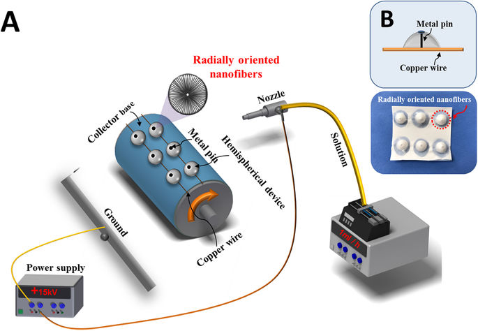
In native cornea, the collagen fibers are arranged in such a way that they radiate from the center of the cornea. Electrospinning may also be used to produce fibers that are radiating from a center. Kim et al (2018) used a non-conducting hemispherical device with a conducting pin in the middle and a wire that forms a ring at the base of the hemispherical device. This setup encourages the formation of a three-dimensional (3D) scaffold with electrospun fibers radiating from the center to the circular wire at the base. Due to the fiber organization, this scaffold was found to exhibit high transparency which is similar to native cornea in the visible wavelength when wetted in PBS.
In cornea treatment, researchers have also looked into other areas where electrospun scaffold may play a part. Ortega et al (2013) investigated the use of constructed electrospun niches to house limbal stem cells which play an important role in cornea regeneration. To construct these artificial niches, electrospinning was used in combination with microfabrication. Microstereolithography via a layer-by-layer photocuring was used to create a template which electrospun poly(lactic-co-glycolic acid) 50:50 was deposited. The template was in the form of a ring with micro-pockets in its circumstance. The deposited electrospun membrane which took on the shape of the underlying template was peeled off the template as the final scaffold.
The ease of loading electrospun fibers with active ingredients has made them highly attractive as drug release agents. Mirzaeei et al (2018) electrospun fibrous meshes of chitosan/polyvinyl pyrrolidone (PVP), chitosan/PVP/polyvinyl alcohol (PVA) and zein/Eudragit loaded with triamcinolone acetonide for the purpose of ocular delivery. Of these, chitosan/PVP electrospun fibers showed a sustained release up to 4 days while zein/Eudragit fibers released 77% of the drugs within this period. Investigation into the release kinetics of electrospun fibrous meshes of triamcinolone acetonide loaded chitosan/polyvinyl pyrrolidone (PVP) electrospun nanofibers showed the preferred zero-order kinetic with constant release rate and independent drug concentration while other polymer combinations adhere to Higuchi model. Grimaudo et al (2020) constructed an ophthalmic insert composed of hyaluronan (HA)/polyvinylpyrrolidone (PVP) nanofibers for the dual delivery of an antioxidant (ferulic acid, FA) and an antimicrobial peptide (ε-polylysine, ε-PL). ε-PL is a naturally produced cationic polyamide which is water-soluble, biodegradable and GRAS food antimicrobial compound active against bacteria, fungi, and yeast. In vitro release tests showed that HA/PVP electrospun fibers loaded with FA and ε-PL completely released both active compounds in 30 min. The presence of ε-PL in electrospun HA/PVP membrane were also shown to inhibit
Pseudomonas aeruginosa and Staphylococcus aureus after 24 h of incubation. Interestingly, a solution of ε-PL with the same concentration as the electrospun membrane did not show inhibition effect on both Pseudomonas aeruginosa and Staphylococcus aureus. While the reason for this is unclear, it may be possible that rapid diffusion of ε-PL solution through the agar gel may have a dilution effect such that the concentration is below the inhibition threshold. Garkal et al (2022) demonstrated the use of thin lutein-loaded electrospun polycaprolactone (PCL) membrane for injection into the eye for treatment of age-related macular degeneration. Lutein was first blended into PCL solution and electrospun to form a thin membrane. In vitro tests showed sustained release of lutein from PCL nanofibers over a 2 months period. The lutein/PCL membrane was found to be stable over a 6 month period, maintaining a drug content of more than 98% after 6 months. However, when the membrane is exposed to light, the drug content drops slightly to 96% after 6 months. Garkal et al (2022) showed that the thin membrane rolled into a tube may be inserted into a 21 Gauge needle from the back and ejected through the tip using a suitable thin wire to push the membrane through. Using a rat model, the membrane was successfully injected into the vitreous of the rat using the needle.
Electrospun conductive nanofibers network with high transparency has been used as an antenna in a soft, smart contact lens for real-time detection of the cortisol concentration in tears [Ku et al 2020]. This antenna needs to occupy a large area over the soft contact lens and exhibit a low sheet resistance for wireless operation of standardized NFC chips. To construct such an antenna, a suspension of Ag nanoparticle ink in ethylene glycol was electrospun to form a network of continuous Ag nanofibers. Thermal annealing was carried out to form a conductive Ag nanofibers network. Finer Ag nanowires were subsequently electrosprayed over the Ag nanofibers network to improve the conductivity of the antenna. The constructed Ag nanofibers/ Ag nanowire antenna has an average sheet resistance(Rs) of 0.3 ohm per square and a transparency of 71% at 550 nm. To maintain conductivity of the antenna over a substantial length of time, passivation by coating the antenna with a layer of parylene elastomeric cover may be used to retard Ag oxidation. The complete device with graphene field-effect transistor as the cortisol immunosensor was tested in vivo using live rabbit and human pilot experiment. The results showed good biocompatibility and reliability of the wireless and battery-free operation of the smart lens.
Published date: 22 September 2020
Last updated: 30 October 2023
▼ Reference
-
Deshpande P, Ramachandran C, Sangwan VS, Macneil S. Cultivation of limbal epithelial cells on electrospun poly (lactide-co-glycolide) scaffolds for delivery to the cornea. Methods Mol Biol. 2013;1014:179.
-
Garkal A, Bangar P, Mehta T. Thin-film nanofibers for treatment of age-related macular degeneration. OpenNano 2022; 8: 100098.
Open Access
-
Kim J I, Kim J Y, Park C H. Fabrication of transparent hemispherical 3D nanofibrous scaffolds with radially aligned patterns via a novel electrospinning method. Scientific Reports 2018; 8: 3424.
Open Access
-
Grimaudo M A, Concheiro A, Alvarez-Lorenzo C. Crosslinked Hyaluronan Electrospun Nanofibers for Ferulic Acid Ocular Delivery. Pharmaceutics. 2020; 12(3): 274.
https://www.ncbi.nlm.nih.gov/pmc/articles/PMC7151120/
-
Kong B, Sun W, Chen G, Tang S, Li M, Shao Z, Mi S. Tissue-engineered cornea constructed with compressed collagen and laser-perforated electrospun mat. Scientific Reports 2017; 7: 970.
Open Access
-
Ku M, Kim J, Won J E, Kang W, Park Y G, Park J, Lee J H, Cheon J, Lee H H, Park J U. Smart, soft contact lens for wireless immunosensing of cortisol. Science Advances 2020; 6: eabb2891.
Open Access
-
Mirzaeei S, Berenjian K, Khazaei R. Preparation of the Potential Ocular Inserts by Electrospinning Method to Achieve the Prolong Release Profile of Triamcinolone Acetonide. Adv Pharm Bull. 2018 Mar; 8(1): 21.
Open Access
-
Ortega I, Ryan A J, Deshpande P, MacNeil S, Claeyssens F. Combined microfabrication and electrospinning to produce 3-D architectures for corneal repair. Acta Biomaterialia 2013; 9: 5511.
-
Wu Z, Kong B, Liu R, Sun W, Mi S. Engineering of Corneal Tissue through an Aligned PVA/Collagen Composite Nanofibrous Electrospun Scaffold. Nanomaterials 2018; 8: 124.
Open Access
▲ Close list