Shape memory material is an important class of "smart" material. This term describes the ability of a material to recover to a pre-determined shape under an appropriate stimulus. Shape memory material has already been used commercially in applications such as orthodontic treatment (Nickel Titanium shape memory wire), stent and in aircrafts (Boeing). Temperature is one of the most commonly used stimuli although material response to other stimulus such as water and light can be found.
Electrospinning has been used to construct nanofiber membrane with shape memory material and shape memory composite with the fibers as filler. A composite made out of electrospun poly-e-caprolactone embedded within a silicone rubber (Sylard 184) matrix has demonstrated strain fixing/recovery of more than 97% for 3 cycles [Luo et al 2009]. Biodegradable and shape memory polymer, poly(D,L-lactide-co-trimethylene carbonate) (PDLLA-co-TMC) has been electrospun and its shape memory effect demonstrated with recovery of more than 98%. With the intention for use in bone tissue engineering, the fabricated scaffold was shown to support osteoblast adhesion and proliferation [Bao et al 2014]. In some cases, shape memory polymer such as Nafion cannot be electrospun to form nanofibers without the addition of electrospinnable polymers. Zhang et al (2013) constructed a nanofibrous membrane out of polyethylene oxide and Nafion blend. The resultant membrane was able to memorize five shapes (four temporary and one permanent shape) with shape recovery of more than 70% [Zhang et al 2013]. For materials made out purely of nanofibers, the shape recovery may be faster due to its high specific surface area. Shape recovery of PDLLA-co-TMC nanofibrous membrane and tube has been shown to take less than 1 min [Bao et al 2014].
It is important to understand the parameters that influence the shape memory characteristics of electrospun fibers. Xi et al (2022) showed that for electrospun fibers with a blend of tert-butyl acrylate (tBA)-co-di(ethylene glycol) dimethacrylate (DEGMA) shape memory polymer and polystyrene (PS), the recovery speed of the electrospun membrane is influenced by the fiber diameter. Tg of the polymer blend were found to increase with reduction in fiber diameter. All the electrospun DEGMA/PS membranes exhibited shape memory effect regardless of fiber diameter. Shape fixity as given by the ability of the membrane to remain at its temporary shape after the force was removed and the membrane cooled, was found to range from 65% to 91% with the membrane having the smallest fiber diameter giving the best shape fixity. It is hypothesized that the better shape fixity of membranes with smaller fiber diameter is due to more fiber crossovers sites. All the membranes were able to achieve 100% shape recovery and the membrane with the largest fiber diameter has the fastest shape recovery speed. Faster recovery of membranes with larger fiber diameter has been attributed to the presence of more amorphous domains in the fiber matrix. It may also be due to greater stiffness of membrane with larger diameter fibers that drives the faster movement.
Electrospun 3D scaffolds have been constructed with shape memory property. This is useful in many surgical application where the incision to insert the scaffold may be made smaller than the recovered scaffold size and implant site. Chen et al (2019) constructed a shape memory 3D scaffold by cross-linking a 3D printed scaffold comprising of chopped nanofibers, hyaluronic acid (HA) and polyethylene oxide solution. In a wet state, the 3D scaffold can be rolled into other shapes before freeze drying to fix the shape. When the scaffold absorbed water, it will return to its original shape in 30s or less.
Table 1. Temperature activated shape memory electrospun material
| Material |
Recovery |
Temperature |
Reference |
| Electrospun poly-e-caprolactone nanofibers in a silicone rubber |
> 97% |
80 oC |
Luo et al 2009 |
| Nafion with 1wt% polyethylene oxide |
> 70% |
65 oC, 90 oC, 110 oC, 135 oC, 160 oC |
Zhang et al 2013 |
| Poly(D,L-lactide-co-trimethylene carbonate) |
> 98% |
39 oC |
Bao et al 2014 |
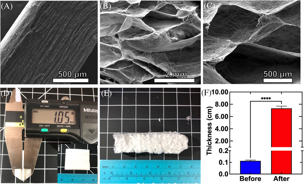
McCarthy et al (2021) also used NaBH4 as a foaming agent for expansion of electrospun polycaprolactone (PCL) nanofiber mats. The resultant PCL matrices were subsequently gelatin-coated and crosslinked to ensure elasticity and matrix shape memory. The foaming agent was so effective that the nanofiber mat with an initial mean thickness of 0.105 cm was expanded to a thickness of 7.33 cm. The gelatin coating and cross-linking significantly enhances the matrix's recovery ability. Coated matrices were able to recover almost 96% of their original shape after manual compression of about 90% while uncoated matrices only recovered to 52% of their original length. In body-simulated fluid (SBF), the gelatin-coated 3D nanofiber matrices were able to recover to 81% of its originally expanded length in 16 seconds while uncoated matrices were only able to recover to 42%.
Another form of memory especially for membranes is to impart strain at its secondary shape. Shape recovery is achieved by releasing the strain from the scaffold. Gunderson et al (2024) demonstrated this by electrospinning and depositing polyglutamic acid-containing polyurethane (PGlu PUr) on a rotating drum collector. With increasing collector rotation speed up to 1000 rpm, there is a corresponding increase in shape memory. This is probably due to greater recoverable strain. Manual programming can be achieved by heating the membrane to 90°C, stretching and cooling under strain. The programmed membrane was able to recover to the original length within 10s when heated to 60°C.
Published date: 19 Feb 2014
Last updated: 29 July 2025
▼ Reference
-
Bao M, Lou X, Zhou Q, Dong W, Yuan H, Zhang Y. Electrospun Biomimetic Fibrous Scaffold from Shape Memory Polymer of PDLLA-co-TMC for Bone Tissue Engineering. ACS Appl. Mater. Interfaces 2014. In press
-
Chen W, Xu Y, Liu Y, Wang Z, Li Y, Jiang G, Mo X, Zhou G. Three-dimensional printed electrospun fiber-based scaffold for cartilage regeneration. Materials & Design 2019; 179: 107886.
Open Access
-
Gunderson A, Ramezani M, Orado T K, Monroe M B B. Programming-via-spinning: Electrospun shape memory polymer fibers with simultaneous fabrication and programming. Smart Materials in Medicine 2024; 5: 477.
https://www.sciencedirect.com/science/article/pii/S2590183424000498 Open Access.
-
Luo X, Mather P T. Preparation and Characterization of Shape Memory Elastomeric Composites. Macromolecules 2009; 42: 7251.
-
McCarthy A, Saldana, L, McGoldrick D, John J V, Kuss M, Chen S, Duan B, Carlson M A, Xie J. Large-scale synthesis of compressible and re-expandable three-dimensional nanofiber matrices. Nano Select. 2021; 1- 14.
Open Access
-
Xi J, Shahab S, Mirzaeifar R. Qualifying the contribution of fiber diameter on the acrylate-based electrospun shape memory polymer nano/microfiber properties. RSC Adv. 2022: 12: 29162.
Open Access
-
Zhang F, Zhang Z, Liu Y, Lu H, Leng J. The quintuple-shape memory effect in electrospun nanofiber membranes. Smart Mater. Struct. 2013; 22: 085020.
▲ Close list