The physical form of organs and tissues are highly varied and tailored to meet their functional objective. While electrospun scaffolds have been widely tested in laboratory on their influence in cell behavior, most cells do not penetrate into the inner depths of the membrane. Such form may be appropriate for studying two-dimensional organs and tissues such as skin and connective tissues but most others like bone and fat tissues takes on a more three-dimensional structure.
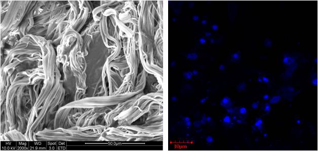
The difference in cell penetration into electrospun 2D scaffolds and electrospun 3D scaffolds were demonstrated by Chen et al (2024). Chen et al (2024) used gas foaming technique to open up the pores between the fibers without excessive application of force. With this, electrospun poly(L-lactide-co-ε-caprolactone) (PLCL)/silk fibroin (SF) nanofiber membrane was expanded to form a 3D scaffold. SEM images of the expanded membrane showed distinct but layered nanofibers with negligible changes in the nanofiber diameters. Inter-fiber gaps increase from 5.63 µm in the pre-foaming membrane to 49.81 µm in the expanded scaffold. Pore sizes also increased from 18.19 µm
2 to 47.37 µm
2 for the expanded scaffold. With this, the density of the scaffolds dropped from 0.175 g/cm
3 in the pre-foaming membrane to 0.033 g/cm
3 in the expanded 3D scaffold and the porosity increased from 51.41% to 77.37%. Chondrocytes cultured on both electrospun 2D membrane and expanded 3D scaffold showed infiltration depth of 83.79 µm in the expanded 3D scaffold compared to just 19.27 µm in the 2D membrane.
Numerous studies on other substrates have provided evidence that cell behavior on two-dimensional substrate and three-dimensional scaffold differs with the later phenotypic cell expression resembling in vivo behavior [Baker et al 2012].
In a study by Lu et al (2023), scaffolds were prepared by co-electrospinning of human umbilical cord mesenchymal stem cells (HUCMSCs) and core-shell fibers (P(AGD)) with the shell made of poly(3-hydroxybutyrate-co-4-hydroxybutyrate) (P34HB) and the core loaded with aqueous solution containing osteogenic compounds L-ascorbic acid-2-phosphate magnesium (ASP), β-glycerophosphate sodium (GP) and dexamethasone (DEX) such that the cells were evenly distributed throughout the P(AGD) scaffold (P(AGD)-CES). In vivo study by subcutaneous implantation of P(AGD)-CES scaffold in rabbit was compared with scaffolds prepared with in vitro co-culturing of HUCMSCs on P(AGD), P34HB nanofiber scaffold but in osteogenic media (P-(ADG)) and P34HB, for 2 weeks prior to implantation. After 8 weeks, the P(AGD)-CES in rabbits showed significant new bone formation and better results compared to the other scaffolds where HUCMSCs were cultured on the scaffolds prior to implantation. The better performance of P(AGD)-CES may be attributed to better distribution of cells throughout the depth of the scaffold compared to the others where the cells were mainly found on the surface.
Electrospun nonwoven membrane with its overlapping fibers are known to restrict cell migration into the scaffold although there are some cases where cells are able to penetrate into the deeper layers. Apart from the fibers in the nanometer diameter, electrospun scaffold is attractive for tissue engineering because it is thought to give the cells a three-dimensional environment. However, from a cell perspective, two or three-dimensionality depends on points of contact between the cell and the scaffold. If the points of contact are multi-directional, the cell is likely to experience a three-dimensional spatial environment. Where the points of contact are from a single side, the cell is likely to treat the scaffold as planar. A scaffold may be thick but does not allow cell penetration or if cell-substrate contact is uni-directional, the environment is likely to stimulate a two-dimensional response. Study by Li et al (2013) using MG-63 osteoblast cells showed that the presence of a three-dimensional environment is more important than the size of the fibers. Their electrospun polycaprolactone (PCL) nanofiber scaffold restricted cell growth on the surface but the microfibrous scaffold allows cell migration into its interior which allows the cells to experience a three-dimensional environment. Osteogenic activity of the cells demonstrated significantly higher activity in the microfiber scaffold than nanofiber scaffold. The most notable advantage of a highly porous, three-dimensional electrospun scaffold is that cells are able to migrate easily into its interior. A three-dimensional space provides a larger spatial environment for cell proliferation and several studies have shown greater cell proliferation on three-dimensional electrospun scaffold compared to two-dimensional scaffold [Blakeney et al 2011, Cai et al 2013, Lee et al 2011]. INS-1 (832/13) cells showed significantly greater proliferation on three-dimensional electrospun scaffold compared to two-dimensional scaffold [Blakeney et al 2011]. Fibroblast cultured on a three-dimensional fluffy scaffold was also shown to adopt a spherical and three-dimensional morphology with much higher level of proliferation (nearly 5 times) than on a two-dimensional membrane after 7 days of culture [Cai et al 2013].
The effect of dimensionality on cell response goes beyond proliferation. An in vitro study by Luong et al on two-dimensional and three-dimensional poly-L-lactide/collagen nanofibrous scaffold found significant difference in mesenchymal stem cell (MSC) response. Contrary to other studies, cell proliferation on the two-dimensional scaffold is significantly greater than three-dimensional (cultured in osteogenic medium). However, bone mineral deposits on three-dimensional scaffold by MSC were significantly greater and denser than the deposits on two-dimensional scaffold after 14 days [Luong 2012]. However, this may be due to the microstructural differences in the 3D scaffold. The microstructure of the 3D scaffold from Luong et al [2012] consisted of bundles of entangled nanofibrous yarn while the 3D scaffold from most others were made of strands of nanofibers. Further, stem cells undergoing differentiation are known to exhibit slower proliferation. Instead of bundles of entangled nanofibrous yarn forming a 3D fibrous structure, Poologasundarampillai et al (2014) constructed a fluffy 3D fibrous scaffold comprising of isolated, randomly organized bioactive glasses fibers. Culturing MC3T3-E1 preosteoblast cell line on this scaffold showed that it is able to induce mineral deposition in the absence of osteogenic differentiation supplements such as ascorbic acid and dexamethasone. Bulysheva et al (2012) using a co-culture of Human telomerase reverse transcriptase (hTERT)-immortalized human foreskin fibroblasts (BJ-hTERT) and hTERT-immortalized HFK-398 human foreskin keratinocytes on a three-dimensional scaffold constructed by low temperature/cryogenic electrospinning showing cell infiltration and high expression of keratin and involucrin which suggest terminal differentiation of the epithelial cells.
3D nanoyarn scaffold has been shown to demonstrate several advantages, at least over 2D membranes. L929 mouse fibroblast was shown to exhibit organized morphology along the nanoyarns with considerable infiltration. Cell proliferation was also found to be significantly greater than 2D membrane after 7 days of culture [Wu et al 2012].
Table 1. hMSC has shown significant osteogenic expression when cultured on a 3D nanofibrous scaffold compared to a 2D nanofibrous substrate [Luong et al 2012].
| 3D nanofibrous scaffold
|
2D nanofibrous scaffold
|
| Cells formed 3D shape or curved around the fiber bundle
|
Cells are flattened on the surface
|
| Slower proliferation rate
|
Higher proliferation rate
|
| Significant ALP expression over 2D scaffold at day 14 and day 21
|
Less ALP expression
|
| Significant OPN expression over 2D scaffold at day 14. OPN expression at 21 days is not significant compared to 2D scaffold
|
Less OPN expression at 14 days
|
| Significant WNT5A expression over 2D scaffold at day 14. WNT5A expression at 21 days is not significant compared to 2D scaffold
|
Less WNT5A expression at 14 days
|
| Significant OCN expression over 2D scaffold at day 14. OCN expression at 21 days is not significant compared to 2D scaffold
|
Less OCN expression at 14 days
|
| Significantly more production of minerals on day 14 compared to 2D scaffold
|
Only a few minerals are produced
|
| Large bone aggregrates with size up to 50 um were observed on day 21
|
Smaller bone aggregates up to 10 um
|
| Ca:P ratio of 1.763
|
Ca:P ratio of 1.402
|
Organization of endothelial progenitor cells (EPCs) has also been shown to be influenced by three-dimensionality of electrospun scaffold. Hong et al (2015) constructed a 3D electrospun scaffold by stacking three layers of nanofibrous membrane with average pore size of 48.21 µm. Membrane with such large pore size was formed over a 0.5 cm diameter hole of an aluminium sheet by electrospinning. This is much larger than the average pore size (<7 µm) of electrospun membrane from a typical flat plate collector. When EPCs are distributed throughout the 3D nanofibrous polycaprolactone (PCL) scaffold, they start to organize into numerous tubular structures after a day. However, in a 2D PCL scaffold, there are no sign of EPCs organizing into tubular structure.
The organization of the electrospun fibers that form the 3D scaffold will also influence cell behaviour. Yang et al (2019) investigated the use of electrospun polycaprolactone (PCL) as fibre-guiding scaffold for periodontal ligament regeneration. In their study, aligned nanofibers and random nanofibers mesh were tested and gelatin was used for binding several mesh layers together to form a three-dimensional scaffold. Human periodontal ligament mesenchyme cells (PDLSCs) were seeded on both scaffolds and examined for their performance in periodontal ligament regeneration. PDLSCs cultured on aligned PCL 3D nanofiber scaffold showed greater alignment and expresses significantly higher level of periostin when compared with the blank control and random 3D nanofiber scaffold. In vivo study was carried out on a Sprague-Dawley rat periodontal fenestration defect model. At 6 weeks, the aligned 3D scaffold demonstrated much more obvious bone formation at the fenestration defect compared to random 3D scaffold and blank control.
Composition of a 3D scaffold would also influence cell behaviour, especially when it is an assembly of different materials. Kumar et al (2019) used an assembly of electrospun aligned poly(ε-caprolactone) (PCL) fiber membrane and collagen get to create 3D composite scaffold. Under static conditions, cultured human mesenchymal stem cells (hMSC) migrated away from the electrospun membrane and into the gel. However, when a mechanical cyclic loading is applied, the cells mostly adhered to the membrane and were aligned in the direction of the fibers. Such behaviour may be due to difference in the stiffness between the gel and the fibers. When load is applied, the cells may preferentially adhered to a stiffer material.
Large pore size in a scaffold poses a challenge in cell seeding. Prior to cell adhesion, the cells are typically rounded up for seeding and they easily fell through the pores in the 3D scaffold. To overcome this challenge, Zaiss et al (2016) constructed a 3D melt electrospun PCL scaffold comprising of highly ordered fibers with large pore size (between 250 to 300 µm) between the fibers and randomly oriented fibers at one end with smaller pore size (between 20 to 80 µm) to prevent cells from falling through. No difference was observed between the osteoblasts cell layer on the large-pores section and the smaller pores section of the scaffold. Melt electrospinning has allowed highly precise deposition of fibers. 3D structures with higher ordered pore sizes can be constructed. However, it is a challenge to get the fiber diameter down to the nanometer range. With melt electrospun structure comprising of fiber with much larger diameter, there may not be significant observations in cellular behaviour. Fuchs et al (2019) compared the response of human osteoblast-like cell line (MG63 cells) on 3D box-shaped scaffolds with pore sizes between 225?µm and 500?µm made from melt electrospun fibers. However, there are no clear pore size preferences from the cells although all of them showed good cytocompatibility according to cell viability, protein concentration, and cell number. It is important to note that the melt electrospun fibers have a diameter of 20µm which is much larger than conventional electrospun fibers. Further tests based on nanometer diameter fibers will be needed to determine the influence of large pore size on osteoblasts.
An alternative to using 3D fluffy nanofibrous block in tissue regeneration is to use chopped electrospun fibers in hydrogel to form a 3D hyrodgel/nanofiber scaffold. This method uses conventional electrospinning to form aligned nanofibers. The aligned nanofibers are subsequently chopped perpendicular to their direction of orientation such that short strand fibers are collected. The chopped fibers are mixed into a hydrogel for implantation or injection. A study by Rivet et al (2015) using chopped electrospun PLLA/fibronectin fibers in hydrogel composed 1.5% SeaPrep (Lonza) agarose and 7.0% Methocel (Dow Chemical) methylcellulose showed extensive astrocyte and macrophage/microglia infiltration into the 3D hydrogel/nanofiber scaffold of a rat model. However, more studies are necessary to determine whether there are any benefits of having nanofibers in hydrogel for tissue regeneration. Instead of a 3D block scaffold made of long continuous fibers, Xu et al (2015) used short strands polycaprolactone (PCL) electrospun fibers from mechanical grounding of deposited PCL fibers followed by suspension in water agglomeration at 55 °C and freeze drying (TISA). A comparison was made with PCL 3D nanofibrous structure made from thermally induced phase separation and porogen leaching (TIPSP) for chondrogenic and osteogenic differentiation of bone marrow mesenchymal stem cells (BMSCs). In osteogenic differentiation, greater ALP activity at 10 days and expression of Runx 2 and BSP after 3 weeks was found in TIPSP scaffold. However, calcium content in TISA scaffold was greater at 3 weeks. At day 4, the expression of Runx2 and Sox9 was greater on TISA compared to TIPSP with the addition of rhBMP2 stimulating a much greater expression of Sox9 (chondrogenic expression) in TISA. This showed that TISA is better able to promote BMP2-induced chondrogenic differentiation of BMSCs.
Preparation of nanofibers suspension for freeze drying to form a 3D scaffold may be easily achieved using a homogeniser. Liu et al (2025) electrospun a blend of gelatin (Gel), polylactic acid (PLA), and magnesium oxide nanoparticles (MgO) solution to form Gel/PLA/MgO fibers. The electrospun Gel/PLA/MgO fibers were added into tert-butanol and homogenised at high speed to form a suspension which was poured into a mold and freeze-dried. Crosslinking was carried out at 180°C for 2h to form the final aerogel scaffolds. The aerogel scaffolds tested on a rat full thickness diabetic wound model showed excellent healing with recovery of epidermal tissue and regeneration of hair follicles and hair by day 21.
In most electrospinning setup, it is a challenge to construct an ordered 3D structure with large pore sizes. Conventional 3D printing using a melt writing method can easily form 3D structures with large pore sizes but the struts are typically in the tens to hundreds of micrometres diameter. Yang et al (2020) tested the combination of three-dimensional printing and electrospinning in the construction of abdominal wall scaffolds. Synthesized polylactic-co-caprolactone (PLC) copolymer was used in 3D printing while collagen was electrospun onto the printed layers. 3D printing (3DP) and electrospinning (ESP) was carried out alternately to create layers of electrospun fibers sandwiched between 3D-printed layers. Samples of 3DP/ESP biocomposite scaffolds and 3DP scaffolds were tested in a rat full-thickness abdominal wall defect model for biocompatibiliity and efficiency. Incorporation of host tissue into the scaffold was greater for the biocomposite group compared to the 3DP group in the first 2 few weeks. This is probably due to the presence of electrospun collagen fibers which offer better cell adhesion. At the end of 12 weeks, integration with the host tissue was also stronger for the biocomposite group than 3DP group as evident from higher tensile strength.
A potential clinical application of three-dimensional electrospun scaffold is for its use as a tissue carrier in fat transplant. It is widely known that the survival of a graft containing cells depends on the ability for nutrient and oxygen transfer to reach the cells within. Otherwise, the cells at the core of the graft will undergo necrosis if angiogenesis is not fast enough. A preliminary study using minced fats mixed with three-dimensional electrospun scaffold have shown that cells were still viable after 4 weeks of culture while cells in at the core of intact fat was no longer viable [Panneerselvan et al 2013]. Further, endothelial cells were also found to be viable in the tissue scaffold mixture and this may potentially accelerate the angiogenesis process. Xu et al (2014) did a comparison of electrospun three-dimensional structure, electrospun two-dimensional structure and commercial 3D scaffold. The 3D electrospun scaffold with adipose derived mesenchymal stem cells (ADMSC) demonstrate better distribution of cells and proliferation compared to two-dimensional scaffold. Investigation of adipogenic differentiation through Oil red O staining also showed more newly secreted fat by ADMSC on the 3D fibrous scaffold followed by 2D scaffold and the commercial 3D scaffold being the worst.
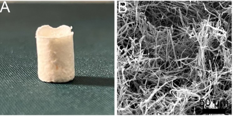
The similarity in the basic structural component of electrospun scaffold with natural extracellular matrix (both being nanofibers) have seen numerous study on the interaction between cells and substrate on electrospun membrane. However, there is much less studies of cell behavior on three-dimensional block nanofibrous scaffold. While fabrication three-dimensional block nanofibrous structure has been a challenge, there are currently several techniques developed to produce such constructs. This presents an opportunity to discover and further our understanding on the behavior of cells in a truly three-dimensional fibrous matrix and applying this knowledge in clinical setting.
Published date: 22 October 2013
Last updated:30 June 2026
▼ Reference
-
Baker B M, Chen C S. Deconstructing the third dimension - how 3D culture microenvironments alter cellular cues. J Cell Sci. 2012; 125: 3015.
Open Access
-
Blakeney B A, Tambralli A, Anderson J M, Andukuri A, Lim D J, Dean D R, Jun H W (2011) Cell infiltration and growth in a low density, uncompressed three-dimensional electrospun nanofibrous scaffold. Biomaterials 32 pp. 1583.
Open Access
-
Bulysheva A A, Bowlin G L, Klingelhutz A J, Yeudall W A. Low-temperature Electrospun Silk Scaffold for In Vitro Mucosal Modeling. J Biomed Mater Res A 2012; 100: 757.
Open Access
-
Cai S, Xu H, Jiang Q, Yang Y. Novel 3D Electrospun Scaffolds with Fibers Oriented Randomly and Evenly in Three Dimensions to Closely Mimic the Unique Architectures of Extracellular Matrices in Soft Tissues: Fabrication and Mechanism Study. Langmiur 2013; 29: 2311.
Open Access
-
Chen Y, Xu W, Pan Z, Li B, Mo X, Li Y, Wang J, Wang Y, Wei Z, Chen Y, Han Z, Lin C, Liu Y, Ye X, Yu J. Three-dimensional gas-foamed scaffolds decorated with metal phenolic networks for cartilage regeneration. Materials Today Bio 2024; 29: 101249.
https://www.sciencedirect.com/science/article/pii/S2590006424003107 Open Access
-
Fuchs A, Youssef A, Seher A, Hochleitner G, Dalton P D, Hartmann S, Brands R C, Muller-Richter U D A, Linz C. Medical-grade polycaprolactone scaffolds made by melt electrospinning writing for oral bone regeneration - a pilot study in vitro. BMC Oral Health 2019; 19: 28.
Open Access
-
Hong J K, Bang J Y, Xu G, Lee J H, Kim Y J, Lee H J, Kim H S, Kwon S M. Thickness-controllable electrospun fibers promote tubular structure formation by endothelial progenitor cells. International Journal of Nanomedicine 2015; 10: 1189.
Open Access
-
Kumar D, Cain S A, Bosworth L A. Effect of Topography and Physical Stimulus on hMSC Phenotype Using a 3D In Vitro Model. Nanomaterials 2019; 9(4): 522.
Open Access
-
Lee J B, Jeong S I, Bae M S, Yang D H, Hei D N, Kim C H, Alsberg E, Kwon I K. Highly Porous Electrospun Nano?bers Enhanced by Ultrasonication for Improved Cellular In?ltration. Tissue Engineering A 2011; 17: 2695.
-
Li T T, Ebert K, Vogel J, Groth T. Comparative studies on osteogenic potential of micro- and nanofibre scaffolds prepared by electrospinning of poly(e-caprolactone). Progress in Biomaterials 2013; 2: 13.
Open Access
-
Liu M, Chen Y, Zhang Y, Zhuang P, Wang J. Breathable functional aerogel dressings facilitate the healing of diabetic wounds. Biomedical Technology 2025; 9: 100071.
https://www.sciencedirect.com/science/article/pii/S2949723X25000030 Open Access.
-
Lu T, Yang L, Li Z, Liu Y, Xu S, Ye C. Immediate implantation of ultrafine fiber slow-release system based on cell electrospinning to induce osteogenesis of mesenchymal stem cells. Regen Biomater. 2023;11: rbad113.
Open Access
-
Luong N T H. Engineered Poly(L-lactic acid)-based nanofibers for osteogenic differentiation of human and mesenchymal stem cells. PhD Thesis. NUS 2012.
Open Access
-
Luong N T H, Liao S, Chan C K, Ramakrishna S. Enhanced osteogenic differentiation with 3D electrospun nanofibrous scaffold. Nanomedicine 2012; 10: 1561
-
Panneerselvan A, Nguyen L T, Su Y, Teo W E, Liao S, Ramakrishna S, Chan C W. Cell viability and angiogenic potential of a bioartificial adipose substitute. J Tissue Eng Regen Med. Ahead of print
-
Poologasundarampillai G, Wang D, Li S, Nakamura J, Bradley R, Lee P D, Stevens M M, McPhail D S, Kasuga T, Jones J R. Cotton-wool-like bioactive glasses for bone regeneration. Acta Biomaterialia 2014; 10: 3733.
-
Rivet C J, Zhou K, Gilbert R J, Finkelstein D I, Forsythe J S. Cell infiltration into a 3D electrospun fiber and hydrogel hybrid scaffold implanted in the brain. Biomatter 2015; 5: e1005527.
Open Access
-
Wu J, Liu S, He L, Wang H, He C, Fan C, Mo X. Electrospun nanoyarn scaffold and its application in tissue engineering. Materials Letters 2012; 89: 146.
-
Xu H, Cai S, Sellers A, Yang Y. Intrinsically water-stable electrospun three-dimensional ultrafine fibrous soy protein scaffolds for soft tissue engineering using adipose derived mesenchymal stem cells. RSC Adv. 2014; 4: 15451.
-
Xu T, Miszuk J M, Zhao Y, Sun H, Fong H. Electrospun Polycaprolactone 3D Nanofi brous Scaffold with Interconnected and Hierarchically Structured Pores for Bone Tissue Engineering. Adv. Healthcare Mater. 2015; 4: 2238.
-
Yang M, Gao X, Shen Z, Shi X, Lin Z. Gelatin-assisted conglutination of aligned polycaprolactone nanofilms into a multilayered fibre-guiding scaffold for periodontal ligament regeneration. RSC Adv. 2019; 9: 507.
Open Access
-
Yang Z, Song Z, Nie X, Guo K, Gu Y. A smart scaffold composed of three-dimensional printing and electrospinning techniques and its application in rat abdominal wall defects. Stem Cell Res Ther 2020; 11: 533.
Open Access
-
Zaiss S, Brown T D, Reichert J C, Berner A. Poly(ε-caprolactone) Scaffolds Fabricated by Melt Electrospinning for Bone Tissue Engineering. Materials 2016; 9: 232.
Open Access
▲ Close list