Home | About | Contact


Microbial exclusion and inhibition using Electrospun fibers

Membrane and coating with microbial exclusion and inhibition property are useful for numerous applications such as filtration, wound dressing and prevention of biofilm formation. Electrospinning is a very simple nanofiber production process which can be used to produce standalone nonwoven nanofibrous membrane or for coating a substrate or surface with nanofibers. Its flexibility in terms of materials that can be used with the process has led to the development of nanofibrous membranes with anti-microbial properties. However, the protective mechanisms against microbes are more than inhibition from material chemistry or additives, the nonwoven nanofibrous network also act as a physical barrier against microbe entry. A recent study also suggested that fiber diameter have an impact on bacteria attachment, proliferation and growth [Abrigo et al 2015].

The most common method of protection against microbes using electrospun fibers is through chemical means. Natural microbial inhibiting materials such as chitin and chitosan and its derivatives may be electrospun into fibers [Seyam et al 2012; Nawalakhe et al 2012; Jung et al 2012]. Inorganic materials such as TiO2 also exhibited anti-microbial capability especially when it is exposed to infrared red light [Aboelzahab et al 2012]. Electrospun cyanoethyl chitosan [Seyam et al 2012] and iminochitosan [Nawalakhe et al 2012] was found to be effective against Escherichia coli (Gram negative), Pseudomonas aeruginosa (Gram negative), Staphylococcus aureus (Gram positive) and Bacillius subtitis (Gram negative). The study by Seyam et al (2012) showed that beaded cyanoethyl chitosan is less effective than smooth fibers in the inhibition of the bacteria. The inhibition ability of chitosan derivatives is dependent on the amount of contact between the bacteria and the material [Seyam et al 2012; Nawalakhe et al 2012]. The mechanism for microbial inhibition by chitosan and its derivatives is based on the interaction between the positively charged chitosan molecules and the negatively charged microbial cell membranes [Goy et al 2009]. Therefore, greater contact between the bacteria and the material will result in more disruption to the bacteria membrane surface and causing its death. For beaded fibers, the points of contacts are less than smooth fibers thus it is unable to reach the level of contact to kill the bacteria. Inorganic materials such as TiO2 work by releasing highly active radical oxygen species when exposed to suitable light wavelength which causes cell and microbial necrosis. This makes it a good material for prevention of biofilm formation. However, in applications such as wound healing, prolonged activation and release of active radical may result in collateral damage to the host cells. Short activation duration may be preferred to reduce cell damage. Dicastillo et al (2018) used Atomic Layer Deposition (ALD) to deposit TiO2 on electrospun polyvinyl alcohol (PVA) template for the construction of TiO2 nanotubes. Following deposition of TiO2 on PVA nanofibers, the PVA template was removed by sintering at 600 °C to produce TiO2 nanotubes. The resultant TiO2 nanotube membrane was tested under dynamic contact conditions against Listeria innocua and Staphylococcus aureus as Gram-positive bacteria and Escherichia coli as Gram-negative bacteria, in accordance to International Normative ASTM E2149-10 with some modifications. When the TiO2 nanotubes concentration (in sterile buffer) was between 150 and 400 µg/mL, the bacteria inhibition under UVA irradiation was high and sometimes total. It is important to note that without TiO2 nanotubes, UVA irradiation inhibited bacteria negligibly, with only 0.1 log cycles reduction. An advantage of using naturally microbes inhibiting material over releasing agent is that the effect of the former is long lasting and does not run out over time.


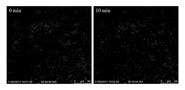
Confocal images of the bacterial colonies in pure TiO2 suspension in S. aureus broth at different times after activation by IR laser for 30 s. Live cells are identified by green pixels and the dead cells by red pixels. [Aboelzahab et al 2012. ISRN Orthopedics, vol. 2012, Article ID 763806, 8 pages, 2012. doi: 10.5402/2012/763806. This work is licensed under a Creative Commons Attribution 3.0 Unported License.]

Electrospun fibers have been used in air and water filtration for removal of particles and have been demonstrated to be very effective for this purpose. Similar, small pore size of electrospun fibrous membrane may also be effective as a barrier against bacteria entry. Lev et al (2012) tested the performance of electrospun polyurethane nanofibers on polypropylene substrate for removal of E. coli from water. At higher nanofibrous layer weight of 3.8 g/m2, exclusion of the bacteria was found to be comparable with commercial water treatment membranes. Chaudhary et al (2014) used an electrospun polyacrylonitrile-silver composite filter media to cover a nutrient media in room condition and passes ambient air through the filter media. When compared to the negative control which is without the protective filter media, the nutrient media protected by the nanofibrous filter remains free of bacteria growth after two months while the unprotected nutrient media show microorganism growth. For application such as wound dressing and food packaging, electrospun nanofibers mesh will be just as effective in keeping bacteria away.

In a recent study, the size of the fiber diameter was found to have some influence on bacteria adhesion and proliferation. When the fiber diameter is close to the size of the bacteria, proliferation was found to be the highest across the bacteria studied (Escherichia coli, Pseudomonas aeruginosa and Staphylococcus aureus) [Abrigo et al 2015]. For rod shaped bacteria such as E. coli and P. aeruginosa, fiber diameters smaller than the bacterial length were found to induce cell death as it attempts to wrap round each fiber. However, the effect of fiber diameter on round S. aureus was less.

Published date: 05 May 2015
Last updated: 24 April 2018

Comments
[+]


Google Ad.