Home | About | Contact


Material Influence on Osteogenic Expression and cell proliferation (Bone)

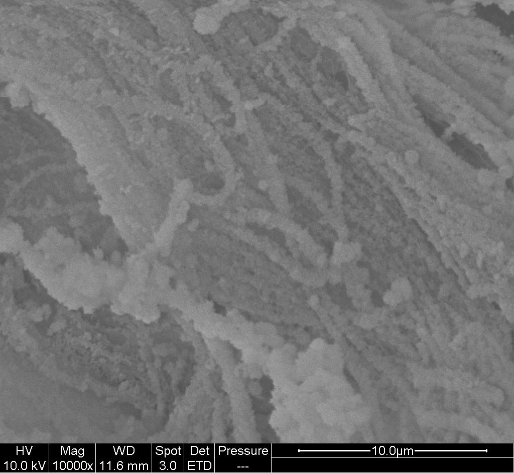
Mineralized nanofibrous scaffold

Nanofibrous topography has been shown to promote mineral deposition and/or osteoblastic expression in osteoblast and mesenchymal stem cells. ALP expression and calcium phosphate mineralization by MSCs isolated from male Wistar rats cultured in osteogenic medium, was found to be significantly greater on PCL nanofiber (dia. 372 nm) membrane compared to smooth PCL film at week 3 [Ruckh et al 2010]. Although most studies suggest that cells are not able to penetrate into the inner depth of electrospun membrane without any modification to the membrane, Yoshimoto et al showed that MSC was able to penetrate into electrospun polycaprolactone (PCL) membrane with evidence of calcification associated with collagen bundles throughout after four weeks of culture in osteogenic differentiation medium [Yoshimoto et al 2003]. Facilitation of differentiation towards osteoblast by nanofiber substrate is not restricted to bone derived mesenchymal stem cells, Polineni et al (2014) showed that adipose derived mesenchymal stem cells (adMSC) showed greater cell proliferation and differentiation when they were cultured on poly-L-lactic-co-glycolic acid (PLGA) nanofiber scaffolds (about 700 nm) compared to PLGA film. Differentiation in the osteogenic medium was further enhanced with the addition of Demineralized Bone Matrix to the PLGA nanofibers.

The material which the scaffolds are made of also has an influence on the proliferation and bioactivities. In particular, natural polymer alone or added to form electrospun fibers have been shown to promote proliferation and osteogenic expression [Venugopal 2007, Prabhakaran et al 2009, Ekaputra et al 2009, Bao et al 2010]. Study by Ekaputra et al of pig bone marrow derived mesenchymal cells showed expression of collagen type I, osteopontin and osteonectin with calcium deposition on PCL/collagen nanofibrous scaffold after 4 weeks of culture in osteogenic media. However, osteocalcin, osteonectin and calcium depositions were not observed on PCL nanofibrous scaffold [Ekaputra et al 2009].

Hydroxyapatite is a major component in bone and it has been hypothesized that its presence on scaffold may stimulate greater osteoblastic activity. Nano-hydroxyapatite may be added to the scaffold either by surface mineralization where the minerals are found on the surface of the scaffold or by mixing into the polymer matrix. Both techniques have been used in the construction of electrospun-based scaffolds and it is interesting to note that the cell response between the two methods of incorporating nHA minerals is different. Whether the nHA is on the surface or within the polymer matrix, osteoblast viability or proliferation on substrates with nHA performs better than neat polymer [Venugopal 2007, Prabhakaran et al 2009, Kim et al 2005, Ngiam et al 2009, Sui et al 2007].

Several studies have demonstrated that MSCs and osteoblast cultured on nanofibers with nano-hydroxyapatite encapsulated within them showed greater ALP and mineral deposition on the substrate compared with neat polymer [Venugopal 2007, Prabhakaran et al 2009, Kim et al 2005, Ruckh et al 2012]. This effect is more apparent where natural polymer is used or blended with man-made polymer. Mineral deposition by human fetal osteoblast cultured on PCL, PCL/nHA, PCL/nHA/Col (blended) and collagen nanofiber membrane was most significant on PCL/nHA/collagen. Proliferation of hFOB on PCL/nHA/Col and collagen nanofiber membrane was also significantly greater than PCL and PCL/nHA membrane [Venugopal 2007]. A study of human fetal osteoblast cultured on PLLA nanofiber membrane, PLLA/nHA nanofiber (blended) membrane, PLLA/nHA/Col (blended) nanofiber membrane and TCP also demonstrated highest ALP and mineralization on PLLA/nHA/Col nanofiber [Prabhakaran et al 2009]. A similar comparative study of PCL, PCL/nHA, PCL/nHA/Col (blended) and collagen nanofiber membrane was carried out using human mesenchymal stem cell (MSC). MSC proliferation was highest on PCL/nHA/Col (blended) nanofibrous scaffold which is similar to the results using osteoblast. Activation of FAK, a protein tyrosine kinase, was also to be greatest on the tri-component scaffold. The favorable cellular response to the tri-component scaffold may be due to greater adsorption of adhesion molecules such as fibronectin and vitronectin as reported in the same study [Matthew et al 2011].

Nano-hydroxyapatite may come in the form of spherical particles, whiskers and rods. Haider et al [2014] compared obsteoblast response to poly(lactide-co-glycolide) nanofiber embedded with spherical HA nanoparticles and nanorod HA and found that the later HA morphology showed significantly higher cell adhesion, proliferation and osteogenesis performance. It was hypothesized that the difference in obsteoblast response is due to difference in the charges on rod and spherical shape HA particles. A rod shape HA will carry more positively charged particles which favours adsoprtion of negatively charged acidic protein while spherical HA carries more positive charges. Nevertheless, both rod and spherical HA embedded nanofibers perform better than neat poly(lactide-co-glycolide) nanofibers.

Instead of having nHA embedded within the nanofiber matrix, Ngiam et al performed mineralization on PLLA and PLLA/Col electrospun membrane to introduce surface nHA. Using TCP, PLLA, PLLA/nHA, PLLA/Col and PLLA/Col/nHA with human fetal osteoblast cells, TCP showed the highest ALP expression at day 7 with PLLA/Col/nHA being the lowest [Ngiam et al 2009]. This differs from the result reported by Prabhakaran et al where at day 10, PLLA/Col/nHA shows the highest ALP expression. Given that the nanofibers for both studies were about 300 nm, the difference in result is likely due to the location of the nHA. In the study by Ngiam et al, the nHA was found on the surface of the nanofiber while nHA was embedded within the nanofiber for Prabhakaran et al. Result by Meng et al on neat and surface mineralized poly(D,L-lactide-co-glycolide) (PLGA) and PLGA/gelatin electrospun nanofiber showed no significant difference in proliferation and osteogenic expression for human osteosarcoma cell [Meng et al 2013]. Contrary to the result showed by Ngiam et al where osteoblast proliferation on nHA coated membrane is lower than the nanofibrous membrane without nHA, Andiappan et al showed higher mesenchymal stem cell proliferation on nHA coated eri silk fibroin nanofibrous membrane compared to those without [Andiappan et al 2013]. Apart from material and cell difference, the average fiber diameter of the fibers from Andiappan et al was much larger (at about 1 um) compared with those from Ngiam et al.

Most in vitro experiments on osteogenic differentiation of stem cells cultured on a material uses osteogenic medium instead of basal medium and essential medium. The stem cells are normally induced towards osteogenic differentiation due to the presence of osteogenic medium and experimental comparison is based on the extent of osteogenic differentiation on different substrates. In a demonstration on the influence of osteogenic differentiation due to physical structure and material, human mesenchymal stem cells (HMSC) showed greater ALP expression when cultured on electrospun nanofibrous scaffold compared to film in basal medium [Polini et al 2011]. To further verify osteogenic differentiation potential, Polini et al (2011) showed osteoinduction of human mesenchymal stem cells (HMSCs) on electrospun poly-caprolactone (PCL)/beta-tricalcium phosphate (TCP) nanofibrous membrane with both the transcription factor Runx-2 and BSP equally upregulated in basal medium as well as in osteogenic medium. Such observation was not found in PCL/TCP film, PCL/hydroxyapatite nanofiber and PCL/hydroxyapatite film.


The enzyme activity was evaluated at day 7 of cellular culture onto different samples, either NFs and control films, for each chemical composition, under BM (Basal medium) or OM (Osteogenic medium) conditions. A) Both a macroscopic view of the Ø 15 mm disks and an optical microscopic image for each sample are shown. Bar: 200 µm. B) Semi-quantification of ALP staining: ALP mean intensity values from image analysis of digital micrographs, after conversion in gray-scale (0-255 bit). Results are reported as mean ± standard deviation. [Polini el al. PLoS ONE 2011; 6(10): e26211.. This work is licensed under a Creative Commons Attribution 4.0 International.]

Other materials, both man-made and naturally derived, have also been shown to drive stem cell differentiation into osteoblasts without the use of osteogenic media. Kashte et al (2019) showed that electrospun PCL fibers surface coated with graphene oxide {GO) and Cissus quadrangularis (CQ) , a plant extract, was able to drive differentiation of human umbilical cord Wharton's Jelly derived mesenchymal stem cells (hUCMSCs) towards osteoblasts lineage without supplementary osteogenic media or growth factors. Verification for differentiation was by Alizarin Red S staining and Von Kossa staining on the cell cultured scaffold to test for calcium deposits. Comparison was made with cells cultured on TCP, PCL, PCL/GO, PCL/GO/CQ, PCL/Graphene {GP} and PCL/GP/CQ. hUCMSCs cultured on PCL/GO/CQ scaffold showed the highest amount of mineral secreted by day 21 while cells cultured on TCP, PCL and PCL/GO were significantly lower. From this study, the presence of GO and CQ have a synergistic effect on increasing hUCMSCs differentiation towards osteoblast-like cells. MXene which is made of 2-dimension transition metal carbides and carbonitrides, has been examined for use in biomedical applications, such as tissue engineering scaffolds. Lee et al (2022) blended MXene nanoparticles into a solution of poly(L-lactide-co-ε-caprolactone, PLCL) and collagen (Col) and electrospun into PLCL/Col/MXene scaffolds for bone tissue engineering. MC3T3-E1 preosteoblasts cultured on the scaffold without osteogenic medium showed significantly enhanced cellular behavior with increased ALP activity compared to scaffolds without MXene. However, the presence of collagen is also important as the PLCL/MXene combination without collagen showed lower ALP activity than the PLCL/Col combination. Mineralized bone nodules were also found on cells cultured on PLCL/Col/MXene scaffolds after 14 days but no mineralization was observed on PLCL and PLCL/Col samples. The osteogenic effect of MXene on preosteoblasts may be attributed to the functional groups of MXene nanoparticles introducing negative charges and the hydroxy groups of MXene forming hydrogen bonds with serum proteins. Carbon elements present in MXene and the nanofiber topography may also contribute to osteogenic effects. Shabestani et al (2022) tested the effect of dihydroartemisinin (DHART), a herbal extract, loaded in electrospun fibers for osteogenic differentiation of adipose-derived stem cells (hADSCs). In their study, the carrier polymers used was a blend of polycaprolactone (PCL) and rat tail collagen types I (Col) to tailor the hydrophobicity and biodegradation of the fibers. DHART in PCL/Col electrospun fibers showed amorphous structure which may increase its bioavailability. Having DHART loaded in PCL/Col electrospun fibers also slowed the release of DHART such that there is sustained release over 28 days. Comparing the differentiation of hADSCs in osteogenic induction medium, hADSCs cultured on PCL/Col/DHART electrospun fibers with 5 wt% DHART showed high expression of osteoblastic markers and enhanced ALP activity over PCL/Col electrospun fibers. 5 wt% DHART was found to perform slightly better than 10 wt% DHART loading in the electrospun fibers. Therefore, 5 wt% DHART in PCL/Col electrospun fibers has the potential to be used in bone tissue regeneration and engineering.

Bone morphogenic protein is known to promote bone formation and is approved by FDA for certain surgical operations such as spinal fusion. Li et al showed that human bone marrow isolated MSC cultured on electrospun membrane consisting of silk fibroin/polyethylene oxide incorporated with BMP-2 and nano-hydroxyapatite (nHA) resulted in the highest calcium deposition and up-regulation of BMP-2 transcript levels compared with polymer only and polymer with either BMP-2 or nHA incorporated [Li et al 2006].


Published date: 14 October 2013
Last updated: 15 August 2023

Comments
[+]


Google Ad.