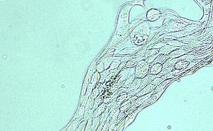
A surface that allows rapid cell adhesion is useful for bedside cell seeding onto scaffold prior to implant or for isolation of cells for diagnostic purpose. A study using osteoblast showed more than 30% cell attachment on the surface of poly-L-lactic acid nanofiber surface compared to about 10% on tissue culture plate (TCP) [Ngiam 2010] at 30 min. Electrospun nanofibrous membrane is known to encourage early cell attachment on its surface. An electrospun membrane may be functionalized to improve cell adhesion rate or made selective towards specific cell type. The smaller pore size of electrospun membranes also gives it the potential for cell separation.
Isolation and in vitro cell expansion is a commonly suggested technique of introducing scaffold with cells. However, this requires two visits to the hospital and a lengthy wait for the cell to multiple to the desired quantity. An alternative is use a scaffold that is able to isolate stem cell from stem-cell rich blood source and implant the stem-cell enriched scaffold. The whole process of blood drawing, isolation and implantation can be completed within one hospital visit. Studies have shown that 25% of hematopoietic stem cell (HSC) was captured on collagen and PLGA/collagen blend in 30 min as compared to only about 2% on TCP[Ma et al 2008]. Significantly higher percentage of MSC was also found to adhere on electrospun membrane at 1 hour (50 to 71%) compared to smooth film (28%) of the same material [Finne-Wistrand et al 2008]. From table 1, it is apparent that a fibrous surface encourages faster cell adhesion compared to smooth surface. Apart from surface roughness, fibers with biomolecules incorporated would further encourage cell adhesion on the substrate [Chan et al 2009].
Table 1. Comparison of cell adhesion/capture on different substrates.
| Reference
|
Cell type
|
Substrate
|
Duration
|
Percentage cell capture/adhesion
|
| Ngiam 2010
|
Osteoblast
|
Poly-L-lactic nanofiber
|
30 min
|
30%
|
| TCP
|
10%
|
| Ma et al 2008
|
Hematopoietic stem cell
|
PLGA/collagen blend (1:1),
|
30 min
|
23%
|
| TCP
|
2%
|
| PLGA
|
8%
|
| Collagen
|
26%
|
| Finne-Wistrand et al 2008
|
MSC
|
Electrospun poly[(L-lactide)-co-(1,5-dioxepan-2-one)]
|
60 min
|
50 to 71%
|
| Film poly[(L-lactide)-co-(1,5-dioxepan-2-one)]
|
28%
|
| Chan et al 2009
|
Human MSC
|
Tissue culture plastic bottom
|
30 min
|
3.2%
|
| Naked glass coverslip
|
3.3%
|
| Gelatin coated coverslip
|
5.3%
|
| Collagen-coated coverslip
|
3.6%
|
| P(LLA-CL) nanofiber, 480 nm
|
9.7%
|
| Air-plasma P(LLA-CL) nanofiber
|
27%
|
| Collagen-coated P(LLA-CL) nanofiber
|
40%
|
| Collagen microfiber, dia. 1140 nm
|
43%
|
| Collagen nanofiber, dia. 390 nm
|
45.1%
|
| CD49a antibody conjugated collagen nanofiber
|
50%
|
| CD29 antibody conjugated collagen nanofiber
|
55%
|
In a novel experiment, mineralized 3D nanofibrous scaffold with yarn microstructure was used to capture nucleated cells from bone marrow. After 20 min, 80% of the nucleated cells were captured on the mineralized 3D scaffold with 76% of the cells expressing MSC markers (CD44) [Ngiam 2010]. It is also possible to use thin nanofibrous membrane or cut nanofibrous membrane to capture the cells prior to meshing the membrane and cells to form a 3D nanofibrous scaffold where structural integrity is not required. Another way of forming 3D scaffold is to stack layers of fiber membrane or rolling the membrane to form a concentric layered cylindrical rod.
The ability to capture and isolate cells may also be used for the capturing of circulating tumor cells. These cancer cells are typically found in small quantities in the blood and the ability to capture it will aid diagnosis. Cao et al (2014) used collagen-blended poly(d,l-lactide-co-glycolide) nanofibers substrate for capturing of K562 leukemia cells. Adhesion of K562 leukemia cells was found to 70% better than tissue culture polystyrene surface with a shorter time. Zhang et al (2012) used electrospun TiO2 functionalized with anti-EpCAM/EpCAM to capture circulating tumor cells from colorectal and gastric cancer patients. Similarly, Liu et al (2014) used MnO2 grafted with anti-EpCAM to capture breast cancer cell line (MCF-7). The nanofibers placed in a microchannel were able to capture 90% of the cells while very few were captured on antibody-grafted glass substrate. Their study also showed that anti-EpCAM grafting contributes to capturing EpCAM-positive cancer cell while EpCAM-negative cancer cell are captured in much lower frequency. Oxalic acid was able to release the captured cancer cell by dissolving the nanofibers with sufficient surviving cells for re-culturing. Zhao et al (2014) used layer by layer technique to functionalize cellulose acetate using a bilayer of poly(diallyldimethylammonium chloride) (PDADMAC) and polyacrylic acid (PAA) through electrostatic interactions. G5.NH2 dendrimer pre-modified with folic acid (FA)and florescein isothiocyanate (FI) was covalently conjugated to the bilayer-assembled nanofibers. With the folic acid, the membrane was able to capture significantly more KB cells (a human epithelial carcinoma cell line) overexpressing FA receptors at 83% after 1 hour compared to the control membrane without surface modifications (38%). This is also significantly more than L929 cells (a mouse fibroblast cell line) and KB cells with low level FA expression at 59% and 66% respectively captured by the surface modified membrane.
Rapid cell adhesion on electrospun fibers may encourage early drug uptakeby the cells. Chu et al (2023) tested the cellular uptake efficiency of released drugs by the nanofibers and nanoparticles using cultured mouse macrophages. At 0.5 h, the cell uptake from the nanofibers was 2.7 times higher than pure nanoparticles although there was no significant difference between the two at 4h. For celecoxib PLGA nanoparticles (Cel-NPs), the cumulative release of celecoxib was 44% within the first 3 hours. Subsequently, a cumulative release of 82% was recorded within 72 h. In contrast, electrospun PVA loaded with Cel-NPs (Cel-NPs-NFs) had a cumulative release of 68% at 168 h which is much slower than Cel-NPs. While drug-loaded nanoparticles showed rapid drug release, the lack of adhesion may delay the drug uptake process by the cells. In contrast, the faster adhesion of the cells on the nanofiber membrane may have allowed earlier absorption of the drugs.
Small pore sizes of electrospun membranes are known to restrict cell penetration. This property may be harnessed in cell separation. Dizon et al (2025) used a combination of electrospinning and vapor-induced phase separation (VIPS) to construct a cellulose acetate (CA) composite membrane for the removal of blood cells from blood-derived solutions. The electrospun CA surface with mean pore size of 2.8 µm was used as the top layer while the VIPS CA layer with mean pore size of less than 0.4 µm was used as the lower layer. The composite was formed by directly electrospinning nanofibers on the VIPS CA membrane layer. The composite membrane was nonhemolytic (hemolysis rate <2%) with a plasma clotting time in the range 16-22 min. Gravity-driven filtration of platelet-poor plasma, platelet-rich plasma, and 10-fold and 5-fold dilutions of whole blood demonstrated complete separation of platelets, red blood cells, and white blood cells from plasma. Further analysis showed that the electrospun layer was able to remove most cells with the VIPS layer removing the remaining cells that penetrated through the top layer. With the VIPS layer alone, rapid cake formation prevented the flow of blood solution through it hence the need for the electrospun layer. However, the composite membrane is not suitable for filtration of undiluted whole blood as the larger cell concentration and subsequent trapping of red blood cells (RBCs) in the depth of the electrospun layer reduces the flow rate resulting in longer filtration duration beyond the plasma clotting time. Given this limitation, the composite membrane may still be useful in the detection of biomarkers in plasma.
Published date: 20 October 15
Last updated: 24 March 2026
▼ Reference
-
Cao X, Kwek K, Chan J K Y, Chan C K H, Lim M. Electrospun nanofibers as a bioadhesive platform for capturing adherent leukemia cells. J Biomed Mater Res A 2014; 102: 523.
-
Chan C K, Liao S, Li B, Lareu R R, Larrick J W, Ramakrishna S, Raghunath M. Early adhesive behavior of bone-marrow-derived mesenchymal stem cells on collagen electrospun fibers. Biomed Mater 2009; 4: 035006.
-
Chu K, Zhu Y, Lu G, Huang S, Yang C, Zheng J, Chen J, Ban J, Jia H, Lu Z. Formation of Hydrophilic Nanofibers from Nanostructural Design in the Co-Encapsulation of Celecoxib through Electrospinning. Pharmaceutics. 2023; 15(3):730.
Open Access
-
Dizon G V, Huang Y J, Lin F C, Maggay I V, Chang Y, Venault A. Assessment of the Blood Separation Performances of Asymmetric Cellulose Acetate Membranes Prepared through Combined Vapor-Induced Phase Separation and Electrospinning. CS Appl. Mater. Interfaces 2025; 17: 31923.
https://pubs.acs.org/doi/full/10.1021/acsami.5c05283 Open Access
-
Finne-Wistrand A, Albertsson A C, Kwon O H, Kawazoe N, Chen G, Kang I K, Hasuda H, Gong J, Ito Y. Resorbable Scaffolds from Three Different Techniques: Electrospun Fabrics, Salt-Leaching Porous Films, and Smooth Flat Surfaces. Macromol. Biosci. 2008; 8: 951.
-
Liu H Q, Yu X L, Cai B, You S J, He Z B, Huang Q Q, Rao L, Li S S, Liu C, Sun W W, Liu W, Guo S S, Zhao X Z. Capture and release of cancer cells using electrospun etchable MnO2 nanofibers integrated in microchannel. Applied Physics Letters 2015; 106: 093703.
-
Ma K, Chan C K, Liao S, Hwang W Y K, Feng Q, Ramakrishna S. Electrospun nanofiber scaffolds for rapid and rich capture of bone marrow-derived hematopoietic stem cells. Biomaterials 2008; 29: 2096.
-
Ngiam M L. Differentiation of bone marrow derived mesenchymal stem cells (BM-MSCs) using engineered nanofiber substrates. PhD thesis. National University of Singapore 2010.
Open Access
-
Zhang N, Deng Y, Tai Q, Cheng B, Zhao L, Shen Q, He R, Hong L, Liu W, Guo S, Liu K, Tseng H R, Xiong B, Zhao X Z. Electrospun TiO2 nanofiber-based cell capture assay for detecting circulating tumor cells from colorectal and gastric cancer patients. Adv. Mater. 2012; 24: 2756.
-
Zhao Y, Zhu X, Liu H, Luo Y, Wang S, Shen M, Zhu M, Shi X. Dendrimer-functionalized electrospun cellulose acetate nanofibers for targeted cancer cell capture applications. J. Mater. Chem. B 2014; 2: 7384.
▲ Close list