Orientation of the fibers is able to facilitate cell migration. Using a radially aligned nanofiber membrane, Xie et al (2010) showed enhanced migration of dural fibroblast towards the center of the membrane compared to membrane with randomly oriented fibers.
Shin et al (2019) used a modified electrospinning setup to create radially patterned polycaprolactone (PCL) nanofibers to encourage faster migration of human bone marrow stem cells from the periphery to the centre of the scaffold. Comparison of cell migration speed was made with randomly oriented electrospun PCL nanofibers. Both radially and randomly oriented nanofibers have a diameter of about 380 nm and interfiber pore size of about 12 nm. From the SEM images of the scaffolds, the radial patterning of the nanofibers are not very clear. However, there are distinct differences in the migration speed of the cells on the radially patterned and randomly oriented nanofibers scaffolds. From day 5 of the cell culture, there is a difference in the cell coverage on the scaffolds. On day 7, the randomly oriented nanofiber scaffold showed a median cell-free area of 84% and the radially patterned nanofibrous scaffold showed a median cell-free area of 49%. This clearly showed the benefits of having radially patterned nanofibers in guiding the cells towards the centre of the scaffold.
Shang et al (2010) showed that rat PDL cells migration on aligned fibers (parallel and cross) was also significantly faster than randomly oriented fibrous scaffold and solvent cast film. Qin et al (2015) reported a detailed study of fibroblast movement on aligned 8 µm diameter polymethylmethacrylate (PMMA) fibers and cast PMMA film. On a fibrillar surface the cells adopted a cytoplasm contraction and expansion mechanism which is similar to that of muscle cells with the nucleus remained highly deformed during migration. On a flat surface, the cells migrate through a sequence of lamellipodia extension, nuclear deformation and retraction of the rear end of the cell. The migration mechanism adopted by the cells on the fibrillar surface allows them to double the contraction amplitude while the contraction of the cells migrating on the flat surface remains unchanged. Similarly, Liu et al (2016) conducted tests on fibroblast movement on aligned 8 µm diameter polymethylmethacrylate (PMMA) fibers and cast PMMA film. Their studies showed that at duration of 24 hour, the migration speed of fibroblasts decreases on a spun cast film while the speed remains constant on aligned fibers. On the 8 µm fiber stacked on one another to form an open 3D structure, the whole surface of the cell adhered and migrate along the fiber surface instead of crossing the space between the fibers.
Cell contraction associated with migration on thin film and 8 µm fiber-(a) Measurement of cell contraction on micron fibers and flat films at day 1 and day 4. Cell contraction on micron fiber is defined as: (cell length at t0- cell length at t1-)/cell length at t1- *: P<0.001. (b) Illustration of cell contraction when migration on flat film. (c) Illustration of cell contraction when migration on 8 µm fibers at day 4 [Qin et al. PLoS ONE 2015; 10: e0119094].
Cell movement on the fibers are sensitive to any disruption to its path. Johnson et al (2009) showed that with human glioma cell that pauses between cell movement often coincides with fiber-fiber intersection even if the angle of misalignment is small (~20 °). Glioma cell migration speed on aligned fibers was much faster than on random fibers with velocity of 4.2 µm/h compared to 0.8 µm/h.
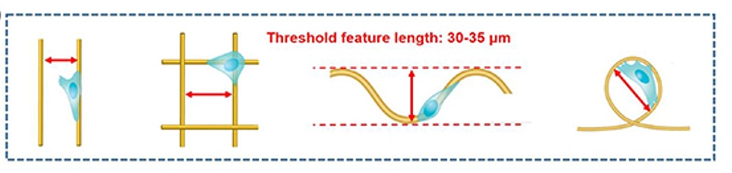
Zhang et al (2022) investigated the migration of human breast cancer adenocarcinoma cell (highest persistent 2D migration among all breast cancer cell lines) on polystyrene-coated glass slides with patterned microfibers (average diameter of 3 + 1 µm) using electrospinning. On parallel fiber tracks, the cells were able to bridge gaps of less than 30 µm between fibers. When the gap is more than 35 µm, the cells would stay on the same fiber. As the width of the cells are below 18 µm, gaps of more than 35 µm, is too great for them to reach across. On grids created by orthogonal overlaid fibers, cells would sometimes cross over to the orthogonal fiber. Consistent with the fibers laid in parallel, bridging between pores formed by the grid only occurred when the gap was less than 35 µm. On other fiber patterns such as wavy and loop, bridging and coverage by the cell is also dependent on whether the gap between segments of the fiber are more than 35 µm or less than 30 µm. The average step speed of the cells were between 0.2 and 0.5 µm min-1 on all four fiber patterns with no significant difference in their speed. It is important to note that the speed of the cell is measured based on the distance covered on the fiber and not a specific direction on the surface.
Schematic diagrams showing that at different fiber patterns, the threshold feature length are all in the range of 30-35 µm [Zhang et al 2022].
Neutrophils are important first responders in an immune reaction as it is able to change shape dramatically to pass through the endothelium layer to get to the damaged or inflamed tissue. However, traditional methods using Boyden chamber to study its migration behaviour may not be accurate as it gives a two-dimensional surface. Jin et al (2015) showed that the smooth surface of the Boyden chamber does not provide sufficient anchorage for neutrophils with the cell adopting a more rounded morphology. In contrast, neutrophils cultured on electrospun fibers attached along the nanofibers and the results are a better representation of cells migration. Using electrospun fibers, they provided further evidence that neutrophils migration is higher in the presence of IL-8 taking into account the effect of gravity.
Stimulation of cell migration on aligned fibers may have clinical relevance in cancer research. Aligned nanofiber is the main component of late-stage breast tumor margin extracellular matrix. Nelson et al (2014) found that breast cancer cells (MDA-MB-231) showed an 82% increase in distance covered in the presence of a CXCL12 gradient on aligned nanofibers while MCF-10A normal mammary epithelial cells showed little response to CXCL12 gradients.
For the same scaffold, a mechanical loading on it as compared to a static scaffold may induce a different migration behavior from the cells. Kumar et al (2019) observed the migration behavior of human mesenchymal stem cells (hMSC) cultured on electrospun aligned poly(ε-caprolactone) (PCL) fiber mesh and electrospun aligned PCL fiber mesh in collagen gel with both scaffolds in static and cyclic loading conditions. In a static fiber/collagen gel scaffold, the hMSCs would migrate into the collagen gel. However, under cyclic loading, most of the cells remain attached to the aligned PCL fiber mesh. Such behaviour may be due to differences in the stfliffness between the gel and the fibers. When load is applied, the cells may preferentially adhered to a stiffer material. hMSCs on aligned fiber mesh for both static and cyclic loaded, and cyclic loaded fiber/collagen gel, the cells were aligned in the direction of the fibers. However, in static fiber/collagen gel scaffold, the cells were less orientated, with actin filaments perpendicular to the underlying fibre direction. Less alignment of the cells under static load may be due to reduced attachment of the cells to the fibers as they migrated into the gel body. The lack of topographical cue on the gel also meant that cells migrated to it have no directional guidance. For the other study groups, the cells were more strongly adhered to the aligned fibers thus the preferential cell alignment with the fibers.
Small inter-fiber pore sizes in electrospun scaffolds typically impedes the migration of cells into it. The inability of cells to squeeze into smaller pore sizes may be due to nuclear stiffness. Heo et al (2020) used Trichostatin A (TSA) for transient softening of the nucleus and showed that it promoted the migration of cells into electrospun scaffolds. The TSA was blended into slow degrading polycaprolactone (PCL) fiber matrix and water soluble polyethylene oxide (PEO) fibers. Without the addition and release of TSA in the cell population, cells seeded atop the fibrous layer remained largely on the surface of the fibers although there was greater infiltration of cells into non-aligned fibers compared to aligned fibers. This is probably due to the greater pore size in non-aligned fibers scaffold. With the electrospun scaffolds releasing TSA, the cells were able to migrate into the fiber network for both aligned and non-aligned fibers and the infiltration depth is the same.
Published date: 28 June 2016
Last updated: 27 September 2022
Heo S J, Song K H, Thakur S, Miller L M, Cao X, Peredo A P, Seiber B N, Qu F, Driscoll T P, Shenoy V B, Lakadamyali M, Burdick J A, Mauck R L. Nuclear softening expedites interstitial cell migration in fibrous networks and dense connective tissues. Science Advances 2020; 6: eaax5083.
Open Access
Jin S W, Park T M, Kum C H, Kim J S, Le B D, Jeong Y H, Kwak J Y, Yoon S. Three-dimensional migration of neutrophils through an electrospun nanofibrous membrane. BioTechniques 2015; 58: 285.
Open Access
Johnson J, Nowicki M O, Lee C H, Chiocca E A, Viapiano M S, Lawler S E, Lannutti J J. Quantitative Analysis of Complex Glioma Cell Migration on Electrospun Polycaprolactone Using Time-Lapse Microscopy. Tissue Engineering C 2009; 15: 531.
Kumar D, Cain S A, Bosworth L A. Effect of Topography and Physical Stimulus on hMSC Phenotype Using a 3D In Vitro Model. Nanomaterials 2019; 9(4): 522.
Open Access
Liu Y, Franco A, Huang L, Clark R, Rafailovich M. Directing Cell Migration by Electrospun Fibers. NSTI-Nanotech 2010; 1: 881.
Nelson M T, Short A, Cole S L, Gross A C, Winter J, Eubank T D, Lannutti J J. Preferential, enhanced breast cancer cell migration on biomimetic electrospun nanofiber 'cell highways'. BMC Cancer 2014; 14: 825.
Open Access
Qin S, Ricotta V, Simon M, Clark RAF, Rafailovich MH. Continual Cell Deformation Induced via Attachment to Oriented Fibers Enhances Fibroblast Cell Migration. PLoS ONE 2015; 10(3): e0119094. doi:10.1371/journal.pone.0119094. Open Access
Shin D, Kim M S, Yang C E, Lee W J, Roh T S, Baek W. Radially patterned polycaprolactone nanofibers as an active wound dressing agent. Arch Plast Surg. 2019; 46(5): 399.
Open Access
Shang S, Yang F, Cheng X, Walboomers X F, Jansen J A. The Effect of Electrospun Fibre Alignment on the Behaviour of Rat Periodontal Ligament Cells. European Cells and Materials 2010; 19: 180.
Open Access
Xie J, Willerth S M, Li X, Macewan M R, Rader A, Sakiyama-Elbert S E, Xia Y. The differentiation of embryonic stem cells seeded on electrospun nanofibers into neural lineages. Biomaterials 2009; 30: 354.
Zhang D, Sheng Y, Piano N, Jakuszeit T, Cozens E J, Dong L, Buell A K, Pollet A, Lei L M, Wang W, Terentjev E, Huang Y Y S. Cancer cell migration on straight, wavy, loop and grid microfibre patterns. 2022 Biofabrication 14 024102.
Open Access