Metal implants are often used in cases where mechanical support is required. Various metals, in particular those with mechanical properties closer to bone, have been investigated for use as implant material. Titanium and its alloy are commonly used as orthopedic or dental implants due to its biocompatibility, good strength and excellent chemical stability. Magnesium alloy is also a potential candidate as its mechanical properties are near to natural bone and it is biodegradable which avoids the need for a second surgical procedure. Gradual degradation of the magnesium alloy will also aid in the smooth transfer of stress to the newly formed bones and facilitate long term recovery of the bone. As with most implants, it is important to foster good integration of the metal implants with the host tissue.
Metal is a commonly used material as collector for electrospun fibers, thus it is relatively straight forward to coat a metal implant with electrospun nanofibers. This has been demonstrated on titanium implant [Ravichandran 2009] and electrospun fibers have also been deposited on magnesium alloy sheet [Soujanya et al 2014] and stainless steel [Khosravi et al 2020]. To enhance integration, mineralization has been carried out on the nanofibers to form polymer-hydroxyapatite composite. Collagen is often used as one of the components in the nanofibers to enhance mineralization [Iafisco et al 2012, Ravichandran 2009]. Ravichandran (2009) showed that adhesion of mesenchymal stem cells (MSC) on titanium plate and alloy is significantly enhanced with nanofiber coating even with man-made polymer such as poly(lactic acid)-co-poly(glycolic acid) (PLGA). Incorporation of the PLGA nanofiber with collagen and hydroxyapatite (HA) further increases the adhesion of MSCs on the implant surface. Titanium alloy coated with PLGA/collagen and HA nanofibers showed better proliferation and exhibited significantly greater alkaline phosphatase activity and mineral secretion than untreated titanium alloy. The relative ease of blending solution for electrospinning has allowed researchers to use multiple additives with selected polymer matrices to create a coating conducive to the intended implant environment. Khosravi et al (2020) investigated the use of electrospun fiber as a coating for 316L stainless steel implants. Poly(ε-caprolactone) (PCL) was selected as the base material to be electrospun due to its known biocompatibility. However, PCL is also known to be hydrophobic hence gelatin was added to reduce its hydrophobicity and to improve cell adhesion. Since this coating is for a bone implant, forsterite (Mg2SiO4) has been added to create a better osteoconductive environment. Forsterite has been known to release Mg ions after implantation which enhances cell proliferation and bone regeneration. In vitro test by immersion of the electrospun PCL/gelatin/forsterite fibers in simulated body fluid (SBF) solution has shown significantly greater hydroxyapatite (HA) formation on the scaffold containing 3 wt% forsterite nanoparticles compared to that with 1 wt% forsterite nanoparticles in both 3 and 7 days.
Coating of 316L stainless steel was also demonstrated by Rezaei et al (2025) with electrospun polyvinyl alcohol (PVA)/Chitosan (60:40 wt. %, respectively)/non-calcined sol-gel-derived bioactive glass (BG-gel) composite fibers. A 15 wt% loading of the BG-gel was found to be optimal as higher loading leads to film formation instead of fibers. In vitro study by immersion of the composite nanofibers in simulated body fluid (SBF) showed hydroxyapatite nucleation on the nanofibers. MG-63 cells cultured on the nanofibers coating exhibited high viability of 93%l after 1 and 3 days of cultivation.
Li et al (2012) went further to load nanofiber with anti-bacterial gentamicin for coating on the titanium implant. The resultant coated implant demonstrated antibacterial efficacy for 1 week against Staphylococcus aureus with significant reduction in adhesion compared with bare titanium implants. The gentamicin-loaded nanofibers did not show any cytotoxicity on osteoblasts. Zhang et al (2014) demonstrated in vitro and in vivo efficacy of vancomycin-loaded poly(lactic-co-glycolic acid) (PLGA) in preventing infection. In vitro showed a drug release profile of an initial burst release on day 1 followed by slow and controlled release over 28 days without any observable cytotoxicity. The drug loaded nanofibrous coating on titanium implant was shown to prevent infection in an animal fracture model where Staphylococci aureus was deliberately introduced to the implant site of the rabbit. In contrast tibia without vancomycin-coated implant showed osteolysis and serious soft-tissue swelling in all animals.
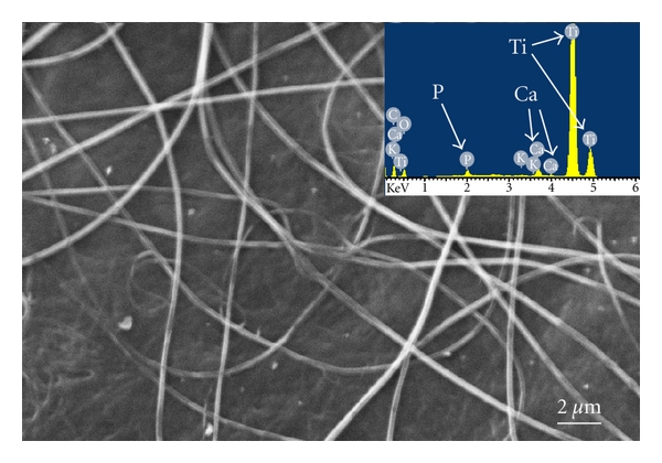
Al-Khatee A et al (2023) used electrospinning to coat the surface of metallic implant with polycaprolactone (PCL)/chitosan/CaTiO3 (CA1) fibers and PCL/chitosan/BaTiO3. The solution for electrospinning was prepared by blending BaTiO3 nanoparticles or CaTiO3 nanoparticles into PCL and chitosan solution. The prepared solution was electrospun onto Ti-25Zr discs and MC3T3-E1 cells were cultured on coated and uncoated discs. Their study found much higher cell viability after seven days on PCL/Chitosan/CaTiO3 (CA1) coated discs at 272% compared to uncoated discs and ALP activity at 5 fold that of uncoated discs. Cell culture on PCL/Chitosan/BaTiO3 (BA1) coated disc compared to uncoated discs had 182% better cell viability and 4 fold increase in ALP activity. The coated discs showed inhibitory activities against Staphylococcus aureus (S. aureus) and Streptococcus mutans (Sterp. mutans) compared to uncoated discs. These antibacterial properties may be due to the presence of chitosan in the fiber. Therefore the nanocomposite fiber coating was able to demonstrate antibacterial properties and encourage cell proliferation and differentiation on metallic implants.
Better cellular response of metal coated with BaTiO3 nanoparticles could be due to its piezoelectric properties. Al-Khateeb et al (2023) blended BaTiO3 and Strontium Titanate (SrTiO3) nanoparticles separately into a mixture of polycaprolactone (PCL) and chitosan (CS) for electrospinning to form a composite coating over acid treated Ti-Zr alloy. SrTiO3 has a high dielectric constant and is mostly found in bone tissue of the human body. The electrospun BaTiO3/PCL/CS coating was found to exhibit dielectric constant close to dry human bone at 100 HZ frequency. Proliferation of MC3T3-E1 pre-osteoblast cells on BaTiO3/PCL/CS and SrTiO3/PCL/CS coated Ti-25Zr plate was found to improve by 125.16% and 111.38% respectively compared to uncoated Ti-25Zr sample.
Having a nanofiber coating on the metal implant has been shown to improve cell adhesion and proliferation on the implant surface and this may potentially foster better integration between the implant and the host tissue. Beside better cell adhesion and proliferation, there are other important requirements such as the ability of the nanofibers to adhere on the surface of the implant without peeling off in vivo. For biodegradable nanofibers, the initial presence of nanofibers may encourage cell adhesion on the implant. However, integration between the host tissue and the implant needs to be investigated once the nanofibers are fully resorbed by the body.
Unlike titanium, magnesium alloys will degrade in physiological condition due to the presence of chloride ions which leads to the formation of magnesium chloride. Without any protection, this will lead to a rapid corrosion and degradation of the magnesium alloy. Electrospinning a layer of polycaprolactone fibers on the magnesium alloy has been shown to significantly reduce its degradation rate by up to five times lower than unprotected samples after 7 days immersion in simulated body fluid [Soujanya et al 2014]. Water contact angle test showed that magnesium alloy is hydrophilic while the alloy sheet with the polycaprolactone coating is hydrophobic. It is likely that the hydrophobicity of the fiber coating prevented or reduces the contact between the fluid and the alloy surface and therefore reduces the corrosion rate. However, based on corrosion protection alone, a PCL dip coating on Mg which fully cover the surface of the metal offered better corrosion resistance than electrospun nanofibrous coating with corrosion rate at 0.3 ng per second for the former and 0.9 ng per second for the latter [Lee et al 2014]. Since electrospun nanofibrous coating is porous, water may still come into contact with the surface of Mg and this will certainly initiate corrosion while the same material completely covered by PCL will only start to corrode after PCL dissolved. For an implant, corrosion resistance is not the only consideration. More tests need to be carried out to determine the effect on cell biocompatibility on the fiber coated magnesium alloy and its impact on corrosion rate.
A potential application of electrospun coated metal is the introduction of infection mitigation property post-surgery. During an open surgery, the wound may be exposed to bacteria which may lead to post-surgery infection. This may be mitigated by having nanofibrous coating on the implant which exhibits antibacterial properties. Aboelzahab et al (2012) electrospun Fe- and Ag-Doped TiO2 nanofibers and test it against Staphylococcus aureus which is a common cause of infection in healthcare settings. Upon exposure of the TiO2 nanofibers and 1 wt% Ag-doped TiO2 nanofibers to infra-red, the survival rate of Staphylococcus aureus was reduced to less than 13% after just 30 s of exposure.
Kiran et al (2018) electrospun polycaprolactone (PCL) nanofibers incorporated with TiO2 nanoparticles on titanium plate surface to increase its bioactivity and imbue antibacterial properties. Their results showed that TiO2 nanoparticles concentration of 7 wt% in PCL nanofibers were toxic to human fetal osteoblastic cell (hFOB). However, at lower concentration of 5 wt% TiO2, proliferation of hFOB cells were enhanced. Antibacterial properties at this concentration was effective against S.aureus with a significant reduction of bacteria to almost 76% under UV light. However, at lower concentration of 2 wt% TiO2, there was insignificant reduction in bacteria.
Published date: 17 June 2014
Last updated: 28 April 2026
▼ Reference
-
Aboelzahab A, Azad A M, Goel V. Necrosis of Staphylococcus aureus by the Electrospun Fe- and Ag-Doped TiO2 Nanofibers. ISRN Orthopedics 2012; 2012: 763806.
Open Access
-
Al-Khatee A, Al-Hassani E S, Jabur A R. Metallic Implant Surface Activation through Electrospinning Coating of Nanocomposite Fiber for Bone Regeneration. International journal of Biomaterial 2023; 1332814.
Open Access
-
Al-Khateeb A, Al-Hassani E S, Jabur A R. Active nanoceramic compound dipped in biopolymers to create composite coating for metallic implant surface. Heliyon 2023; 9: e19594.
Open Access
-
Khosravi F, Khorasani S N, Khalili S, Neisiany R E, Ghomi E R, Ejeian F, Das O, Mohammed H N E. Development of a Highly Proliferated Bilayer Coating on 316L Stainless Steel Implants. Polymers (Basel). 2020; 12(5): 1022.
Open Access
-
Kiran A S K, Kumar T S S , Sanghavi R, Doble M, Ramakrishna S. Antibacterial and Bioactive Surface Modifications of Titanium Implants by PCL/TiO2 Nanocomposite Coatings. Nanomaterials 2018; 8(10): 860.
Open Access
-
Iafisco M, Foltran I, Sabbatini S, Tosi G, Roveri N. Electrospun Nanostructured Fibers of Collagen-Biomimetic Apatite on Titanium Alloy. Bioinorganic Chemistry and Applications 2012; 2012: 123953.
Open Access
-
Lee D H, Park C H, Kim C S. An experimental investigation on corrosion rate of Mg electrode using EQCN and improvement in anti corrosion rate of Mg electrode by surface coating. Digest Journal of Nanomaterials and Biostructures 2014; 9: 1171.
-
Li L L , Wang L M, Xu Y, Lv LX. Preparation of gentamicin-loaded electrospun coating on titanium implants and a study of their properties in vitro. Arch Orthop Trauma Surg 2012; 132: 897.
-
Ravichandran R. Biomimetic Surface Modification of Dental Implant for enhanced Osseointegration. MEng Thesis. National University of Singapore 2009.
Open Access
-
Rezaei M, Rabiee S M, Jamaati R, Rahmani S. Surface modification of AISI 316 L stainless steel with polyvinyl alcohol/chitosan/sol-gel bioactive glass coating by electrospinning method. Applied Surface Science Advances 2025; 28: 100801.
https://www.sciencedirect.com/science/article/pii/S2666523925001096 Open Access.
-
Soujanya G K, Hanas T, Chakrapani V Y, Sunil B R, Kumar T S S. Electrospun Nanofibrous Polymer Coated Magnesium Alloy for Biodegradable Implant Applications. Procedia Materials Science 2014; 5: 817.
Open Access
-
Zhang L, Yan J, Yin Z, Tang C, Guo Y, Li D, Wei B, Xu Y, Gu Q, Wang L. Electrospun vancomycin-loaded coating on titanium implants for the prevention of implant-associated infections. International Journal of Nanomedicine 2014; 9: 3027.
Open Access
▲ Close list