Home | About | Contact


Water vortex

3D nanofibers 3D nanofiber setup
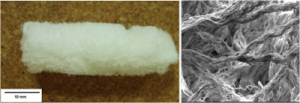
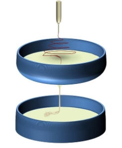
Left: Freeze-dried nanofibrous 3D structure. Right: View of the nanofibrous arrangement under SEM (W.E. Teo et al. Current Nanoscience 2008; 4: 361. © BSP). Schematic of the electrospinning setup

Fabricating a nanofibrous block with comparable length, width and height poses a significant challenge to the electrospinning process. In the electrospinning process, nanofibers are deposited in layers as they settle on top of one another. Due to the nano-dimension, it would take a very long time for the nanofibers to build up to a thick layer. The pore size across the thickness of the nanofibrous membrane is also significantly smaller to the pore sizes as seen from above. Therefore, the electrospinning setup needs to be modified such that the orientation of the nanofibers are not restricted to a single plane.

In this concept, a water vortex is created by having the water draining down a sink hole at the bottom of a basin. This creates a water flow that runs from the surface through a hole before releasing the contents in a reservoir below. When the electrospun fiber is deposited on the water surface, the flow carries the fiber with it at the same time consolidating the mesh to form a yarn as it passes through the sink hole. The consolidated nanofibrous yarn is allowed to fall into a basin of water below the reservoir above as shown in the figure. Within a few minutes, clumps of nanofibrous yarn would be collected in the water basin. These can then be gathered and free-dried to give a block of nanofiber clump.

The freeze dried nanofibrous clump is soft to the touch and is very similar to a ball of cotton wool. Under the SEM, the pore size between the nanofiber yarn can be up to several hundreds of micron. Such a scaffold may be used in tissue engineering. Post fabrication mineralization has been carried out to form a 3D polymer-hydroxyapatite scaffold [Teo 2009] and has been tested for bone regeneration [Ngiam 2010].

Since the dynamic liquid electrospinning is based on modifications of the collector, changes may be made to the electrospinning source without affecting the output form. Guo et al (2016) was able to use this setup to electrospin core-shell nanofibrous yarn 3D scaffold using coaxial nozzle. This is useful in tissue engineering and implantable scaffolds where bioactive agents may be added to it. The resultant scaffold retains large pore sizes typical of 3D scaffold fabricated using this method and was demonstrated to allow fibroblast infiltration [Guo et al 2016].

Instead of allowing the yarn to collect as a clump in the reservoir, a collector may be placed in the falling water to gather the yarn. This may allow some organization of the yarn as it forms the 3D scaffold [Li et al 2012].

Published date: 09 August 2012
Last updated: 14 February 2017

Comments
[+]


Google Ad.