Colorimetric sensor is very useful and convenient for detecting the change agent where accurate quantitative result is not required. This is mostly used for rapid screening on site before further quantitative tests are carried out if necessary. Electrospinning is a simple method for fabricating fibers down to the nanometer scale and its high surface area allows rapid detection of any changes in the environment. There are a few methods of introducing colorimetric changes to electrospun fibers in response to the stimulus. Gold nanoparticles in a suspension or matrix are known to show different colors depending on its dispersion characteristic. Conjugated polymers are known to exhibit changes in properties in response to stimulus.
Gold nanoparticle is a potential candidate for colorimetric sensor due to color changes from its aggregation or dispersion which is easily distinguishable by the naked eye. Its aggregation and dispersion characteristics are influenced by interaction with other molecules such as salt and biomolecules. Based on this concept, Pule et al (2015) used electrospun strips made of polystyrene containing gold nanoparticles for detection of 17β-estradiol. In unspiked effluents, the strips changed from white to brown. However, for effluents spiked with 17β-estradiol, the strips turned pink. At higher concentration of 17β-estradiol, the color developed further to blue. Changes in the strips color were observed within 120s with good sensitivity towards 17β-estradiol. Color change was detectable by the naked eye for concentration of 17β-estradiol as low as 100 ng/ml. Senthamizhan et al (2018) used dithiothreitol capped gold nanocluster (DTT.AuNC) as fluorescent probe for detection of Cu2+. Electrospun porous cellulose acetate fibers (pCAF) were used as protective carrier for DTT.AuNC. Cellulose acetate was first electrospun into fibrous membrane before immobilizing DTT.AuNC on its surface by dipping in a DTT.Au suspension followed by washing. Attachment of DTT.AuNC onto pCAF may be from the hydrogen bonding between the thiol (-SH) or hydroxyl group in DTT with acetate group of CA. It is interesting to note that DTT.AuNC immobilized on pCAF is more stable than DTT.AuNC immobilized on nonporous CAF. Greater stability of DTT.AuNC on pCAF may be due to deeper penetration of DTT.AuNC into the pores of pCAF and the protection of DTT.AuNC within its pores. When tested for detection of Cu2+, there is a visible detectable limit of 1 ppm with the membrane changing from red to blue for DTT.AuNC@pCAF under UV. Sensing response of DTT.AuNC on nonporous CAF to Cu2+ was much weaker. For Hg2+, Cd2+ and Zn2+ at concentration of 20 ppm, there was no change in fluorescence behavior from Hg2+ while enhanced fluorescent intensity due to Zn2+ and Cd2+ was not visible to naked eye under UV light.
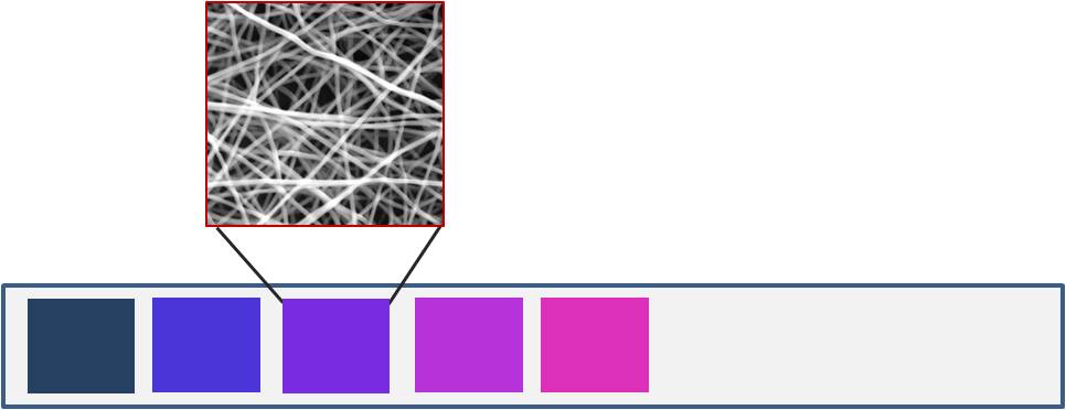
Polydiacetylenes (PDA) are known to change from blue to red upon application of stimulus. This characteristic has been tested for use in the detection of organic solvents. PDA from different diacetylene monomers respond differently to volatile organic solvents (VOC). Yoon et al (2007) was able to use this to create electrospun poly(ethylene oxide) (PEO), and tetraethyl orthosilicate (TEOS) embedded with four PDAs from different diacetylene monomers (10,12-pentacosadiynoic acid (PCDA, CH3(CH2)11CtC-CtC(CH2)8-COOH) (1), 5,7-eicosadiynoic acid (ECDA, CH3(CH2)11CtC-CtC(CH2)3COOH) (2), N-(pyridin-3-yl) pentacosa-10,12-diynamide(PCDA-AP) (3), and 8,10-henicosadiynoic acid (HCDA, CH3-(CH2)9CtC-CtC(CH2)6COOH) (4)). These PDAs were subsequently used for the identification of four different VOCs (chloroform, tetrahydrofuran (THF), ethyl acetate (EA), and n-hexane)by the changes in the color of the strips. Further taking the advantage of polydiacetylene (PDA) and its colorimetric transitions when exposed to certain organic solvents, Yoon et al (2009) narrowed down the DA monomer selection to 10,12-Pentacosadiynoic acid (PCDA)-ABA 1, a derivative of PCDA and aminobutyric acid (ABA), and aniline (AN)-derived DA monomer PCDA-AN 2 to display different colorimetric responses to a variety organic solvents. Their study showed that various compositions of (PCDA)-ABA 1 and PCDA-AN 2 showed different degree of blue-purple-pink transitions. By having different compositions of (PCDA)-ABA 1 and PCDA-AN 2 in a strip, they showed that it is possible to differentiate between the solvents, chloroform, THF, methylene chloride, methanol, acetone, hexane, DMF, AcOH and CH3CN.
Similarly, Ali et al (2016) constructed an electrospun membrane for the detection of adulteration in gasoline using toluene and thinner. Their sensor was made of polycaprolactone (PCL)/polydiacetylenes (PDA) fibers which were constructed by incorporating pentacosadiynoic acid (PCDA) in PCL and electrospinning with post-spinning UV-polymerization. This produces a distinctly blue membrane. In pure gasoline, there are no color changes to the membrane or damage to the fibers. However, when exposed to toluene and thinner vapors, the membrane changes to red within seconds to a couple of minutes. Observation under SEM showed dissolution of the fibers.
Interestingly, Yapor et al (2017) showed that polydiacetylenes (PDAs) coupled with either electrospun poly(ethylene oxide) (PEO) or polyurethane (PU) was able to demonstrated colorimetric changes to the presence of Escherichia coli ATCC25922 bacterial cells. The composite fibers where constructed by blending 10,12-pentacosadiynoic acid (PCDA) with a supporting polymer, poly(ethylene oxide) (PEO) and polyurethane (PU), and the blended solution was electrospun to produce fiber composites. The electrospun fibers were subsequently photopolymerized using UV irradiation to form PEO/PDA and PU/PDA. When the nanofibers membrane was exposed directly to E. coli ATCC25922 grown on Luria-Bertani agar, there was a rapid colorimetric response. The mechanism for the color change has been attributed to endotoxins released by Gram-negative bacterial strain which disrupts the hydrogen bonding within the PDA molecules and causes color change.
1,8-naphthalimide-based colorimetric derivative has been developed and used extensively in fluorescence detection for HTMs such as Hg2+, Cd2+, and Cu2+. Liang et al (2017) tested the effectiveness of a fluorescent chemodosimeter, namely a 1,8-naphthalimide-based colorimetric derivative, 1-benzoyl-3-[2-(2-allyl-1,3-dioxo-2,3-
dihydro-1Hbenzo[de]isoquinolin-6-ylamino)-ethyl]-thiourea (BNPTU), with high selectivity for Hg2+ in aqueous solutions encapsulated in electrospun poly(N-isopropylacrylamide)-co-(N-methylolacrylamide)-co-(acrylic acid) (poly(NIPAAm-co-NMA-
co-AA)) fibers. Fe3O4 nanoparticles were also added to give the membrane magnetic properties. The resultant composite membrane was found to be strongly selective against Hg2+ in a mixture of other common ions displaying a blue emission in the presence of Hg2+ while otherwise staying green. With 10 wt% BNPTU, the composite membrane was very sensitive to Hg2+ with significant blue shift observable at 10-3M Hg2+ concentration.
Dithizone (DTZ), also known as 1,5-diphenylthiocarbazone is an organic compound that forms complexes with many metals. With its high density color, Dewi et al (2023) used this for colorimetric determination of Cr6+. To increase selectivity of the sensor, DTZ was first complexed with Co2+ which would be replaced by Cr6+ in an oxidoreduction reaction. To construct this nanofibrous membrane sensor, DTZ was first mixed into a polyurethane (PU) solution and electrospun into a nanofibrous membrane form. Cobalt nitrate solution was then dripped onto the DTZ/PU membrane to form the DTZ-Co2+ complex. When tested with Cr6+ solution, the color changed from red to magenta in a few seconds and is visible to the naked eye. The DTZ-Co2+/PU nanofibrous membrane showed high sensitivity with a lower limit of detection (LOD) of 0.001 mgL-1 and a wide linearity range (0.001 - 1.0 mgL-1. In comparison, the best formulation of DTZ, PU and Co2+ applied as a microwell plate film showed a lower sensitivity with LOD of 0.018 mg L-1 and a narrower linearity range of 0.01-5.0 mg L-1. Tests with 13 interfering cations, Ca2+, Mg2+, Zn2+, Cu2+, Fe3+, Fe2+, Mn2+, Ni2+, Pb2+, As3+, Se2+, Cd2+ and Hg2+ showed no effects on the colour response to Cr6+hence demonstrating the selectivity sensor.
In the area of oxygen sensing, optical sensors based on the luminescence quenching principle has attracted much interest due to its general excellent sensitivity, accurate detection, low detection limit, fast response/recovery etc. Mao et al (2018) investigated the advantage of having silver nanowire (AgNW) in electrospun palladium octaethylporphine-poly(methyl methacrylate) (AgNWs@PdOEP-PMMA) microfiber mats prepared by electrospinning. In the presence of oxygen, phosphorescence intensities of the AgNWs@PdOEP-PMMA microfiber mats gradually decreased as the oxygen concentration increases from 0 to 100%. This reduction in phosphorescence intensities were attributed to efficient quenching effect of the microfiber mats with its high porosity and interconnected pores when exposed to oxygen.
AgNWs@PdOEP-PMMA-sensing microfiber mats exhibited a swift response (approx. 1.8 s) and improved sensitivity (by 243% for the range of oxygen concentration 0-10% and 235% for the range of oxygen concentration 0-100%) compared to the pure PdOEP-PMMA microfiber mat.
Easy detection of bacteria or bacteria activities are very useful in many applications from food preparation to bacteria contamination in an area. Colorimetric means are particularly attractive due to the convenience of visual cues. Kinyua et al (2022) constructed a colorimetric sensor made of electrospun chitosan/polyethylene oxide nanofibers (CS/PEO NFs) grafted with chromogenic substrate 5-bromo-4-chloro-3-indolyl-β-D-glucuronide (X-Gluc) for the purpose of detecting bacteria, Escherichia coli, activities. In the presence of the enzyme β-glucuronidase (β-GUS) from Escherichia coli (E. coli), hydrolytic cleavage of the chromogenic substrate X-Gluc leading to the release of an indoxyl derivative which forms a blue colored dichlorodibromo indigo dye in air. Comparing the reaction rate of CS/PEO NFs and CS/PEO hydrogel of the same mass and enzyme concentration, the higher surface area of CS/PEO NFs gave a much higher reaction rate and greater amount of dye released. The limit of detection for β-glucuronidase by the nanofibers was 13 nM compared to 25 nM of CS/PEO hydrogel. The lower limit of detection for the CS/PEO NFs was 13 nM compared to 25 nM CS/PEO hydrogel sensors. For the lower limit of quantification for the enzyme, CS/PEO NFs was 45 nM while CS/PEO hydrogel was 60 nM.
Published date: 20 September 2016
Last updated: 10 October 2023
▼ Reference
-
Ali S, Ahmed F, Khatri A, Khatri M. Colorimetric Sensor for Detection of Adulteration in Gasoline using Polydiacetylene Electrospun Fibers. Pak. J. Anal. Environ. Chem. 2016; 17: 38.
-
Dewi I R, Rujiralai T, Putson C, Cheewasedtham W. A novel double metal-dithizone functionalized polyurethane electrospun nanofiber and film for colorimetric determination of hexavalent chromium. RSC Adv. 2023; 13: 2852.
Open Access
-
Kinyua CK, Owino AO, Kaur K, Das D, Karuri NW, Müller M, Schönherr H. Impact of Surface Area on Sensitivity in Autonomously Reporting Sensing Hydrogel Nanomaterials for the Detection of Bacterial Enzymes. Chemosensors. 2022; 10(8):299
Open Access
-
Liang F C, Luo Y L, Kuo C C, Chen B Y, Cho C J, Lin F J, Yu Y Y, Borsali R. Novel Magnet and Thermoresponsive Chemosensory Electrospinning Fluorescent Nanofibers and Their Sensing Capability for Metal Ions. Polymers 2017; 9: 136.
-
Mao Y, Liu Z, Liang L, Zhou Y, Qiao Y, Mei Z, Zhou B, Tian Y. Silver Nanowire-Induced Sensitivity Enhancement of Optical Oxygen Sensors Based on AgNWs-Palladium Octaethylporphine-Poly(methyl methacrylate) Microfiber Mats Prepared by Electrospinning
ACS Omega 2018; 3 (5): 5669.
Open Access
-
Pule B O, Degni S, Torto N. Electrospun fibre colorimetric probe based on gold nanoparticles for on-site detection of 17β-estradiol associated with dairy farming effluents. Water SA 2015; 41: 27.
-
Senthamizhan A, Celebioglu A, Balusamy B, Uyar T. Immobilization of gold nanoclusters inside porous electrospun fibers for selective detection of Cu(II): A strategic approach to shielding pristine performance. Scientific Reports 2018; 5: 15608.
Open Access
-
Yapor J P, Alharby A, Gentry-Weeks C, Reynolds M M, Alam A K M M, Li Y V. Polydiacetylene Nanofiber Composites as a Colorimetric Sensor Responding To Escherichia coli and pH. ACS Omega 2017; 2: 7334.
Open Access
-
Yoon J, Chae S K, Kim J M. Colorimetric Sensors for Volatile Organic Compounds (VOCs) Based on Conjugated Polymer-Embedded Electrospun Fibers. J Am Chem Soc 2007; 129: 3038.
-
Yoon J, Jung Y S, Kim J M. A Combinatorial Approach for Colorimetric Differentiation of Organic Solvents Based on Conjugated Polymer-Embedded Electrospun Fibers. Adv Func Mater 2009; 19: 209.
▲ Close list