Home | About | Contact


Modifying biodegradation rate of electrospun fibers

Biodegradable polymers are attractive for use as implantable scaffolds as the eventual replacement of the scaffold by host tissues will eliminate or reduce any long term foreign body response complications. In drug loaded nanofibers, the degradation rate of the polymer may also influence the release rate of the drugs. The desired degradation rate of the polymers will depend on the intended application and performance of the implant which may range from weeks to years. Degradation rate of different polymers are also varies widely with polyglycolic acid having a half-life of only a few weeks to polycaprolactone having a half-life of many years. There are several methods of altering the degradation rate of a material such as having material mixtures, its physical characteristic, degradation characteristic and post electrospinning treatment.

When selecting a biodegradable material for an intended application, it may be necessary to refine its rate of degradation to bring it closer to the desired rate. A simple method is to use a mixture of materials with some components that degrades at a faster rate or influence the degradation of the primary material. Spagnuolo and Liu (2012) used a blend of two biodegradable L-tyrosine-based polyurethanes material but with different degradation rates to control the degradation of the electrospun scaffold. Similarly, Liu et al (2008) was able to show reduced mass reduction when they increase the ratio of slow degrading polycaprolactone to faster degrading PLGA in blended electrospun nanofibers. This concept has also been used by other materials mixture to control the degradation rate [Zhang et al 2011]. Thao et al (2021) electrospun a mixture of small intestinal submucosa (SIS) and poly(ε-caprolactone-ran-l-lactide) (PCLA) to control the degradation rate of the electrospun nanofiber membrane for drug release. Electrospun SIS membrane easily solubilizes in biological media hence it is not suitable for use as drug release carrier for extended drug release. PCLA on the other hand is hydrophobic and the membrane from electrospun mixture of SIS and PCLA showed a gradual increase in hydrophobicity as the ratio of PCLA increases. Degradation test using collagenase showed the SIS sheet completely degraded after 1 day, electrospun SIS/PCLA (5:1) membrane showed 50% degradation in a day but remained the same for 3 days and electrospun PCLA membrane showed no degradation. For a membrane made of SIS and PCLA, the initial fast degradation is likely due to the degradation of SIS and the degradation rate slows down significantly due to slow degradation of the remaining PCLA. The different material characteristics may influence drug release rate if multiple drugs of different properties are loaded. Thao et al (2021) loaded dexamethasone (Dex) and silver sulfadiazine (AgS) into electrospun SIS/PCLA (5:1) membrane. In an in vivo drug release test, Dex, which is more water soluble than AgS, saw a rapid release on the first 3 days (40%) followed by linear release of the remaining amount over 14 days. For AgS, 37% was released on the first 3 days with little release over 21 days. The more water soluble Dex may have greater affinity towards SIS in the polymer mixture which has a higher degradation rate. Since AgS is poorly water soluble, more of it may get trapped in PCLA and the slow degradation of PCLA would have slowed its release after the initial burst release.

Degradation rate of a material may also be tailored by having additives that catalyst its degradation or removal of catalyst. Kim et al (2003) showed that it is possible to control the rate of degradation for non-woven poly(d,l-lactide) (PDLA) nanofiber scaffolds by blending PDLA with other materials such as poly(lactide-co-glycolide) (PLGA) random copolymers, poly(lactide-b-ethylene glycol-b-lactide) (PLA-b-PEG-b-PLA) triblock copolymers, and a lactide (used as a hydrolytic catalyst). In this mixture, PLGA and PLA-b-PEG-b-PLA are used as bridging material to control the hydrophicity of the membrane and the retention of lactide to affect the overall degradation rate. For a mixture comprising of 40wt% of PDLA, 15 wt% of lactide (used as a catalyst), 20 wt% of PLA-b-PEG-b-PLA triblock copolymer and 25 wt% of low molecular weight PLGA(LA/GA=50/50), a weight loss of 65% was recorded which is much greater than pure PDLA.

In contrast to adding a catalyst to increase the degradation rate of PLA, several studies have showed that the addition of nano-hydroxyapatite (nHA) into electrospun poly(L-lactide) (PLLA) fibers is able to reduce its degradation rate [Mansourizadeh et al 2014; Zhao et al 2006]. The presence of nHA probably neutralizes the acidic degraded products from PLLA and this reduces its degradation rate compared to pure PLLA [Zhao et al 2006]. However, for a hydrophobic material, the presence of nHA may increase its hydrophilicity and this may in turn increase its degradation rate.

Adjusting the hydrophobicity of a polymer is another way of influencing its degradation rate. Polycaprolactone (PCL) is known to exhibit a very slow degradation rate even in its nanofibrous form. Gaharwar et al (2014) were able to significantly accelerate its rate of degradation by introducing nanoclay into its matrix. The nano-clay facilitates adsorption of water into the nanofiber matrix and this contributes to the hydrolytic degradation of the PCL chains. With 10% nanoclay, the thinner fibers were completely degraded in 24 h. Degradation of the PCL fibers was through surface erosion as seen by the gradual reduction of fiber diameter. This also applies to the addition of nHA to a hydrophobic material. Xu et al (2007) showed that with small amount of nHA (3 wt%) grafted onto poly(L-lactide) (PLLA), the degradation rate is slower than pure PLLA. However at higher amount of nHA, the degradation rate is faster than pure PLLA. This has been attributed to the increased presence of nHA on the surface of the fibers which reduced its hydrophobicity. Niu et al (2017) used blending to load electrospun poly(L-lactic acid) (PLLA) nanofibrous scaffold with amorphous calcium phosphate nanoparticles (ACP). Similar to the results from Xu et al (2007) who used nHA, the degradation rate of the ACP loaded PLLA nanofibers showed faster degradation rate with 5wt% and 10wt% ACP. This has been attributed to increased hydrophilicity which facilitate faster hydrolytic degradation. Further, comparing the morphology of the fibers between pure PLLA fibers and ACP loaded fibers, degradation of the former showed continued smooth surface while the latter showed microbumps and holes. The microbumps and holes may be due to dissolution of ACP into the solution and its presence increases its surface area for faster degradation.

The physical form of the scaffold has an impact on its degradation rate. Spagnuolo and Liu (2012) showed that the degradation of electrospun nanofibers for L-tyrosine-based polyurethanes is faster than the corresponding film of the same composition. This is likely to be due to the greater surface area of electrospun nanofibers. Similarly, Choi et al (2004) showed that the degradation of electrospun poly(3-hydroxybutyrate-co-3-hydroxyvalerate) was faster for smaller diameter fibers and slowest for film. However, degradation rate of PLGA has been shown to be slower in its electrospun form compared to film [Shang et al 2010]. Presence of catalytic by-products trapped within a block material may increase its degradation rate compared to highly porous nanofibers where by-products are quickly released into the media as in the case of PLGA. Natu et al (2012) did a long term (30 months) comparison of the degradation rate of PCL material produced by different methods, electrospinning, solvent casting, melt compression and supercritical fluid processing. Their result showed that in the first year, degradation rate was consistently slowest for electrospun fibers compared to the materials produced by the other three methods. However, at 30 months, the molecular weight of the electrospun nanofibers was almost the same as the materials produced using other methods.

Beyond considering the degradation rate of the polymer, there may be other requirements such as materials availability, cost, manufacturability, compatibility with additives and others. Therefore, it may sometimes be necessary to tailor the degradation rate of the selected polymer rather than selecting the polymer based on its degradation rate.

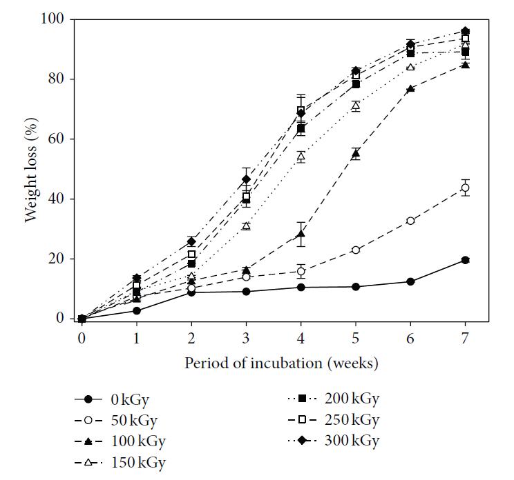
Several post process treatments may be used to change the degradation rate of the polymeric scaffold. Treatment through radiation may cause cross-linking which increases the molecular weight of the polymer or main chain scission which reduces the molecular weight. While the former will increase the half-life of the polymer, the latter reduces it. Lee et al (2015) carried out several tests on the effect of electron beam irradiation dosage on electrospun poly(lactide-co-glycolide) (PLGA). With a dosage of 50 kGy, a molecular weight reduction of more than half was recorded and the corresponding weight loss at seven weeks doubles to 40%. At 100 kGy, the weight loss at seven weeks doubles again to 80%. Increasing irradiation dosage from 100 kGy to 300 kGy progressively increases the weight loss to more than 90% over the same time period. Mechanical properties such as its modulus, tensile stress and elongation at break also deteriorate with increasing E-beam dose. The most pronounced effect is at 150 kGy on the elongation at break where it was reduced from 194% at 100 kGy to just 8%. There is no observable visual difference in the fiber diameter or physical morphology throughout the tested dose from 50 kGy to 300 kGy.

Similarly, exposure of polymer to strong ultra-violet rays (UV) may increase its degradation rate. Dong et al (2008) showed that exposure of poly(D,L-lactic-co-glycolic) acid (PLGA, 75:25) and poly(L-lactide-co-e-caprolactone) [P(LLA-CL) [70:30] nanofibrous meshes to UV for 1 hour reduces their molecular weight by 46% and 35% respectively [Dong et al 2008]. This reduction in molecular weight will certainly cause an increase in its degradation rate.

Weight loss of e-beam irradiated PLGA nanofibers as a function of length of incubation in PBS (37 °C) over a period of 7 weeks. Data are expressed as the mean ± S.D. of 3 samples [Lee et al. Modification of PLGA Nanofibrous Mats by Electron Beam Irradiation for Soft Tissue Regeneration. Journal of Nanomaterials 2015; 2015: 295807. This work is licensed under a Creative Commons Attribution 3.0 Unported License.].

Published date: 16 June 2015
Last updated: 21 December 2021

Comments
[+]


Google Ad.