The most common form of electrospun structure is a 2D membrane or mesh. In electrospinning, a high voltage power is used to stretch the solution from the emitter to be deposited on a flat collector. Numerous in vitro studies have been carried out on this form of electrospun scaffold and many have shown promising results in terms of cell adhesion and proliferation. In terms of tissue repairs, there are many applications where a mesh form may be used.
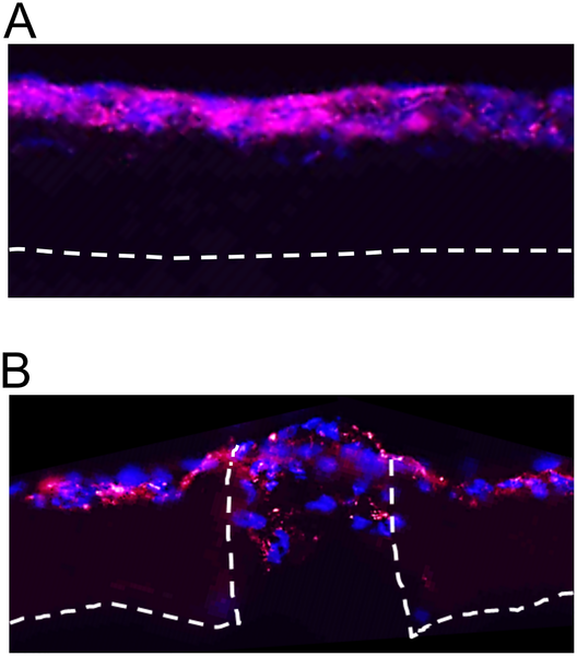
An obvious tissue where a mesh scaffold can be used is the skin. Not surprisingly, there have been extensive studies on the use of electrospun scaffolds as skin grafts. Rho et al (2006) using electrospun collagen scaffold on full thickness wound on Sprague-Dawley showed faster early stage healing (up to 7 days) compared to wound without scaffold. However, no significant difference in the wound closure was found between wound with and without collagen scaffold during the later stage of the study with both groups demonstrating complete epithelialization after 4 weeks. Due to rapid degradation of collagen scaffold, it may not contribute to later stage healing of the wound. A synthetic polymer with longer degradation rate may better facilitate wound healing throughout the duration of the healing process. In a study by Han et al (2007) using Athymic nude mice full-thickness skin wound model, poly(3-hydroxybutyrate-co-3-hydroxyvalerate) (PHB) nanofiber scaffold showed better healing than PHB/collagen. The best result comes from PHB nanofiber scaffold seeded with cocultured (3 - 5 days) dermal sheath (DS) cells and epithelial outer root sheath (ORS) cells. Bonvallet et al (2015) showed that electrospun a 70% collagen I and 30% poly(ε-aprolactone) nanofibers membrane perforated with 160 µm diameter holes and seeded with F344 fibroblasts (35,000 cells on a 15 mm diameter scaffold) performed better than acellular membrane using a syngeneic Fischer 344 (F344) rat full-thickness skin defect model. The presence of the through pores also allowed the cells to migrate into the depth of the scaffold. F344 fibroblasts were isolated from the rat skin to avoid rejection of the implanted cells.
In vitro study showed that extracellular matrix (ECM) was found to be deposited in the 160 µm diameter holes with maximum pore filling by 14 days. Comparing the performance of the scaffolds where cells have been cultured for 1 day and 4 days, the scaffold with longer cell cultivation prior to implantation showed better recovery. This could be due to a combination of higher cell density and presence of more ECM on the scaffold.
In the treatment of bone defects or injury, the scaffold to be used is not always in the form of a block scaffold. In some cases, a mesh form is preferred as guided bone regeneration membrane. These membranes are usually used in calvarial defect, omentum and sterna injury which does not require thick scaffold. The electrospun membrane in this application typically functions as a guided bone regeneration graft which works as a barrier to prevent soft tissue invasion into the defect site [Dimitriou et al 2012].
In cavarial defects repair, a scaffold with a minimum level of stiffness and strength is preferred especially if the defect is larger. Pensa et al (2019) constructed such a scaffold using electrospun poly-epsilon;-caprolactone (PCL) /gelatin and 3D printed poly(lactic acid) (PLA) mesh. Instead of electrospinning and depositing PCL/gelatin fibers on 3D printed mesh, the PLA filaments were 3D printed on the electrospun fiber mesh. This has the advantage of creating a stronger bond between the 3D printed structure and the electrospun mesh. In 3D printing, the extruded filament in its semi-molten state will partially melt the PCL component in the electrospun fibers due to its lower melting point. An in vivo rat cranial defect study showed that the reinforced electrospun scaffolds with the 3D printed struts facing the outer surface of the skull did not elicit an immune or foreign body response after 20 weeks of implementation.
To reduce infection from the surgery, Jin et al (2018) incorporated Ag-CaP into electrospun chitosan (CS) fibers. Ag ion is known to be an effective antibacterial agent. The resultant electrospun Ag-CaP/CS exhibited a sustainable release of Ag+ and inhibited the adhesion and growth of Staphylococcus mutans. Its biocompatibility was shown by the adhesion and proliferation of bone marrow stromal cells cultured on it over 11 days. Cell viability was highest for CaP/CS electrospun scaffold. When there were Ag added (0.075% and 0.14%), the cell viability was reduced which showed that Ag+ does inhibit cell proliferation. However, with the added Ag, cell viability on Ag-CaP/CS electrospun scaffold was still better than pure electrospun CS scaffold.
The pore sizes of electrospun meshes are typically less than 10 µm. This creates a barrier for cells to penetrate through it. This property of electrospun fibers membrane has allowed it to be used as an anti-adhesion barrier film between injured site and the surrounding tissue. This is important for preventing peritendinous adhesion where the injured tendon repair site suffers from fibrotic adhesion which limits the mobility of the joint. Using an electrospun hyaluronic acid-grafted poly(caprolactone) (PCL-g-HA) nanofibrous membrane, Chen et al (2014) showed that PCL-g-HA electrospun membrane is effective in reducing peritendinous adhesion in an animal model. The study also showed that the presence of HA is necessary to reduce fibroblast attachment while the membrane provides a physical barrier for fibrotic adhesion.
The dura mater is a connective tissue membrane that protects the brain tissue. This membrane is made out of various fibrous layers which electrospun mesh can imitate. Chuan et al (2020) investigated the potential use of electrospun poly(lactic acid) stereocomplex (PLAc) with poly(D-lactic acid)-grafted tetracalcium phosphate (g-TTCP) composite nanofiber membrane as dura matter substitute. Of particular importance is the mechanical properties compatibility with native human dura matter. Comparing different ratios of PLAc and g-TTCP, it was found that a ratio of 45:55 electrospun nanofiber membrane after heat treatment at 100 °C for 1 h exhibited mechanical properties closest to native dura matter. The elongation at break and ultimate tensile strength of the fabricated dura matter substitute were 111.2% and 6.46 MPA respectively which is comparable to native dura matter at 116% and 7 MPa respectively. Good biocompatibility of the dura matter substitute was demonstrated by culturing of mesenchymal stem cells (MSC) and its differentiation into neuron-like cells.
Electrospun nanofiber mesh may be used to treat abdominal wall defects. The mesh needs to demonstrate little inflammatory response and its mechanical properties should match the compliance of native tissue to intra-abdominal pressure (IAP). Liu et al (2020) constructed an electrospun polycaprolactone (PCL)/silk fibroin (SF) mesh integrated with amoxicillin (AMX)-incorporating multiwalled carbon nanotubes (MWCNTs). AMX is a broad-spectrum antibiotic which is added to prevent infection of the treatment site. Coating of electrospun PCL/SF mesh with AMX@MWCNTs was carried out through an amidation reaction using 1-ethyl-3-(3-dimethyl aminopropyl) carbodiimide (EDC)/N-hydroxysuccinimide (NHS) solution. The modification of PCL/SF mesh with AMX@MWCNTs increases the elongation at break of PCL/SF mesh from 60% to 150% for PCL/SF/AMX@MWCNTs which is
closer to the expansion of native abdominal wall. PCL/SF/AMX@MWCNTs mesh also demonstrated good in vitro inhibition of E. coli with sustained release over many days. An in vivo rat model with subcutaneous implementation showed that the PCL/SF/AMX@MWCNTs mesh induces less mesh-induced inflammatory and foreign body responses than PCL/SF mesh within 14 days.
Published date: 12 January 2021
Last updated: 04 May 2021
▼ Reference
-
Bonvallet PP, Schultz MJ, Mitchell EH, Bain JL, Culpepper BK, Thomas SJ, Bellis S L. Microporous Dermal-Mimetic Electrospun Scaffolds Pre-Seeded with Fibroblasts Promote Tissue Regeneration in Full-Thickness Skin Wounds. PLoS ONE 2015; 10(3): e0122359. doi:10.1371/journal.pone.0122359.
Open Access
-
Chen S H, Chen C H, Shalumon K T, Chen J P. Preparation and characterization of antiadhesion barrier film from hyaluronic acid-grafted electrospun poly(caprolactone) nanofibrous membranes for prevention of flexor tendon postoperative peritendinous adhesion. International Journal of Nanomedicine 2014; 9: 4079.
Open Access
-
Chuan D, Wang Y, Fan R, Zhou L, Chen H, Xu J, Guo G. Fabrication and Properties of a Biomimetic Dura Matter Substitute Based on Stereocomplex Poly(Lactic Acid) Nanofibers. Int J Nanomedicine 2020; 15: 3729
Open Access
-
Dimitriou R, Mataliotakis G I, Calori G M, Giannoudis P V. The role of barrier membranes for guided bone regeneration and restoration of large bone defects: current experimental and clinical evidence. BMC Medicine 2012; 10: 81.
Open Access
-
Han I, Shim K J, Kim J Y, Im S U, Sung Y K, Kim M, Kang I K, Kim J C. Effect of poly(3-hydroxybutyrate-co-3-hydroxyvalerate) nanofiber matrices cocultured with hair follicular epithelial and dermal cells for biological wound dressing. Artif Organs 2007; 31: 801.
-
Jin S, Li J, Wang J, Jiang J, Zuo Y, Li Y, Yang F. Electrospun silver ion-loaded calcium phosphate/chitosan antibacterial composite fibrous membranes for guided bone regeneration. International Journal of Nanomedicine 2018:13 4591.
Open Access
-
Liu Z, Zhu X, Tang R. Electrospun Scaffold with Sustained Antibacterial and Tissue-Matched Mechanical Properties for Potential Application as Functional Mesh. International Journal of Nanomedicine 2020; 15: 4991.
Open Access
-
Pensa N W, Curry A S, Bonvallet P P, Bellies N F, Rettig K M, Reddy M S, Eberhardt A W, Bellies S L. 3D printed mesh reinforcements enhance the mechanical properties of electrospun scaffolds. Biomater Res 2019; 23: 22.
Open Access
-
Rho K S, Jeong L, Lee G, Seo B M, Park Y J, Hong S D, Roh S, Cho J J, Park W H, Min B M. Electrospinning of collagen nanofibers: effects on the behavior of normal human keratinocytes and early-stage wound healing. Biomaterials 2006; 8: 1452.
▲ Close list