There are many ways to deliver drugs such as by injection, intravenous, implantation and oral. Oral intake is probably the least invasive and most acceptable to patients for drug administration. This is also a very feasible method for treatment along the digestive tract. However, a key challenge is to ensure that the drug is being released at the requisite site. An advantage of electrospun fibers is that their general small diameter and large surface area facilitates rapid drug release. This ensures that the drugs are fully unloaded before the carrier is transported further down the digestive tract.
For mucoadhesive drug delivery where topical application is preferred, ultrafast release of the drug alone is insufficient as the patient may ingest most of them. To facilitate optimal retention of the drug in the area of application, a less soluble backing material may be used [Dott et al 2013; Tyagi et al 2014]. Dott et al (2013) electrospun polyvinyl alcohol (PVA) loaded with citric acid, diphenhydramine and glycerol on a film cast from PVA and hydroxypropylmethylcellulose (HPMC). While the electrospun film alone takes less than 4 s for complete disintegration, cast film with electrospun fiber took about 13 s. Cast film alone with the same components and composition took about the same time for complete disintegration but its mechanical strength is weaker than cast film with electrospun coating.
Topuz et al (2022) electrospin pure HPβCD loaded with Piroxicam (Px), a nonsteroidal anti-inflammatory drug, for rapid sublingual delivery of Px. The solution was prepared in water mixing Px and HPβCD for a day hence there is no use of potentially toxic solvents. Smooth fibers of diameters 500 nm or less were produced from electrospinning the solution with higher loading of Px leading to smaller fiber diameters. Px in the HPβCD fibers were found to form inclusion complexes and the initial highly crystalline Px became amorphous as demonstrated by the absence of sharp peaks in XRD patterns. When placed in artificial saliva, the dissolution of the membrane was completed within a second and about 90% of Px was released within a minute and close to 100% of the Px was released after 10 min. Px in the fibers were found to be stable after six months of storage which can be attributed to the complexation between the CD and Px.
To impart different functionality to the fiber, a core-sheath fiber structure may be used. Although core-sheath fiber structure is more commonly used to control the release rate of drugs, a highly water soluble sheath material will also allow rapid release of the drugs located in the core of the fiber. Yu et al (2011) constructed a core-sheath fiber with the sheath comprising of PVP and sucralose as sweetener and core material comprising of PVP and a drug, acyclovir. In vitro results showed that all acyclovir were released in less than a minute but the release rate of acyclovir powder is much longer with less than fifty percent released after an hour. Slow release rate of acyclovir powder probably due to the size of the particles which is about 100 µm.
In a similar study to Yu et al (2011), Ning et al (2021) used coaxial electrospinning to construct a core-shell fiber with sucralose and low molecular weight polyvinylpyrrolidone (PVP) as the shell and the drug diclofenac sodium (DS) and high molecular weight PVP as the core. In this setup, the shell materials are not electrospinnable on their own while the core high molecular weight PVP is able to electrospin into fibers. Therefore, in this coaxial electrospinning, it is the core solution that carries the electrospinning process while the shell solution coats over the inner material. Sucralose and DS are crystalline in their solid pure forms which increases the time for their dissolution. The presence of PVP and the molecular interaction between sucralose and DS and their respective PVP mixture hinders their ability to crystallize. This coupled with the fast vaporization of the solvent during electrospinning ensures that sucralose and DS remain in their amorphous state after fiber formation. Comparing the rate of dissolution, DS in its powder form took almost an hour for complete dissolution while the core-shell electrospun fibers containing DS took less than 16s to completely disappear. Such rapid dissolution of the electrospun fibers can be attributed to the hydrophilic carrier material, high porosity of the nanofibrous membrane and amorphous state of the drug.
Asawahame et al (2015) investigated the potential use of water-soluble polyvinylpyrrolidone nanofibers loaded with propolis (PVP-propolis) as antibacterial agent in the mouth. Pure PVP-propolis nanofibers were found to be completely wetted in 45s while the addition of Tween 80 and small amount of flavouring agents reduced the wetting time to less than 2s. When tested against Streptococcus mutans (S. mutans), PVP-propolis nanofibers containing 0.6 MIC (minimum inhibitory concentration, 1.172 mg/mL) were effective in decreasing the adherence of the bacteria to the glass surface. The addition of Tween 80 and flavouring agents does not show significant difference in the inhibition of S. mutans adherence to glass compared to fibers without. Sipos et al (2019) demonstrated the potential of aceclofenac-loaded triethanolamine-containing polyvinylpyrrolidone (PVP) for oral release of aceclofenac. In vitro test showed that the drug release from the nanofibrous web is almost immediate, and complete within a minute. In contrast, dissolution rate of the pure drug was 51% at the first minute and 85% at 3 min. Such rapid release of aceclofenac in electrospun PVP membrane is due to the high surface area of nanofibers, amorphous state of the drug and solubility of the polymer in water.
A limitation of current treatment for oral mucosal lesions is that the drug stays for only a short duration which render their topical application ineffective. This has prompted researchers to develop electrospun drug loaded mucoadhesive membranes with longer-lasting adhesive properties [Santocildes-Romero et al 2017, Colley et al 2018]. Colley et al (2018) constructed an electrospun dual-layer mucoadhesive system comprising of an outer electrospun hydrophobic polycaprolactone (PCL) backing layer and an mucoadhesive layer formed by electrospinning polyvinylpyrrolidone (PVP), Eudragit® RS100 and polyethylene oxide (PEO) . The drug, clobetasol-17-propionate, was dissolved in the PCL/Eudragit® RS100/PEO solution before electrospinning. Testing the mucoadhesive patch on human volunteers found that the average residence time of the patch on gingiva, tongue and buccal mucosa was 118, 43 and 96 min respectively. Most of the volunteers responded positively to the adherence of the patches and over 88% of volunteers felt little or no irritation from the patches. In vitro study showed slow sustained of the drug over a 6 h period with 20 % and 50% of the drug released after 30 and 180 mins respectively. In vivo test on mini pig buccal mucosa model for the clobetasol-17-propionate patch showed marked levels of drug was detected in the mucosal epithelium after just 30 min application using the 20 µ g/ml loaded patch but declined significantly by 60 min but remained constant for up to 240 min. Such significant difference in the drug release profile between in vivo and in vitro results highlights the challenges in finding the optimal composition of drug and material selection for mucosal adhesion and drug release.
In paediatric oral drug delivery, having the drug delivered in film form will improve swallowability and favourable mouthfeel. The taste of the drug may also be masked through the encapsulation of the drug within the film matrix. Abdelhakim et al (2021) used co-axial electrospinning to create a core-shell fiber with a model bitter drug, chlorpheniramine maleate blended into the core polymer matrix. It was found that having Kollicoat® Smartseal (KCT) in the core with the drug and Eudragit® E PO (E-EPO) in the shell is the most effective for taste masking. E-EPO electrospins well hence it is able to form an intact and uniform covering over the KCT/chlorpheniramine maleate core and provide effective taste-masking. Further, a higher concentration of E-EPO was used to form the solution for electrospinning compared to KCT which may provide a better protective covering. However, when E-EPO was used as both the core and the shell, the masking effect was less than the use of two different polymers. This could be due to the presence of an interface layer with two different polymers that retards the migration of the drug to the surface. Both E-EPO and KCT are pH sensitive and are insoluble above pH 5.0. At pH 1.2 which mimics gastric conditions, the film released approximately 75% of the drugs within 45 min due to the high surface area of the electrospun fibers. This release rate is in line with the immediate release formulations' guideline of releasing 70% of the drug in that timeframe (Ph. Eur. 5.17.1).
For drugs targeting the gastrointestinal tract, oral delivery is the best way to administer them either in the form of pills or capsules. For microbiome-based drugs, there is an additional challenge of maintaining the viability of the microbes during processing and storage. The most common method of preparing microbiome-based drugs is through freeze drying but this process potentially causes a lot of stress which may lead to loss of bacteria viability. Electrospinning provides an alternative for drying the bacteria in a carrier polymer. Vass et al (2020) investigated the use of water soluble hydroxypropyl-beta-cyclodextrin (HP-β-CD) as the carrier polymer for Clostridium butyricum which is a anaerobic spore-forming bacteria normally found in the human gastrointestinal tract. After electrospinning of HP-β-CD loaded with C. butyricum spores or cells, the loaded fibers were grinded into powdered form for compression into pills. These processes did not lead to a reduction in the bacteria viability. Electrospun sporulated C. butyricum was also found to remain viable after 1 year of aerobic storage at ambient temperature. Sporulated C. butyricum contained 30% bacteria spores and 70% vegetative cells. For electrospun vegetative cells, 1 year of aerobic storage at ambient temperature renders them unviable. Therefore, electrospinning is a promising formulation technology for microbiome delivery applications.
The pH value changes along the digestive tract and this is one condition that can be considered when selecting the material for targeted drug delivery. The stomach gastric fluid has a pH of about 1.2, intestinal fluid about pH 6.8 and colonic fluid about pH 7.4 [Li et al 2018]. Biodegradable pH-sensitive polymers containing ortho ester groups such as D,L-lactide have been shown to increase the drug release rate in acidic media [Qi et al 2008]. Electrospun Eudragit fibers showed very limited release at pH 2.0 but sustained release over approximately 8 h at pH 6.8. This makes it suitable for an oral drug delivery system to target the intestinal tract [Illangakoon et al 2014]
Drug delivery targeted at the lower digestive tract need to pass through the esophagus and the acidic environment of the stomach. The colon is known to harbour abundant colonic microflora population and their corresponding enzyme activities. Understanding this environment, polysaccharides-based microflora-activated colonic delivery system made from chitosan, alginate, and cellulose may be used to protect the drug until it reaches the colon. Li et al (2018) electrospun a solution mixture of pectin-coated salmon calcitonin (SCT) liposomes, sodium alginate (SA) and polyvinyl alcohol (PVA). The intention is to deliver calcitonin to the colon for promoting bone absorption of calcium. In their in vitro tests using simulated gastric fluid containing pepsin (SGF, pH 1.2), simulated intestinal fluid containing trypsin (SIF, pH 6.8) and simulated colonic fluid containing pectinase (SCF, pH 7.4), the release of SCT was found to be 22.6% in SGF, 16.8% in SIF, and 49.6% in SCF, respectively. This demonstrated the feasibility of using electrospun colon-specific fiber mat for bioactive peptide delivery. Another way of delaying the release of drug by the carrier until it reaches the colon is to use a protective barrier that is resistant to dissolution in acid but soluble in pH neutral environment. Yang et al (2018) produced electrospun core-shell fibers with a shellac she'll to protect the drug loaded polyvinylpyrrolidone (PVP) core. As a material, shellac is soluble in neutral condition but insoluble in acid condition. This makes it a suitable protective barrier during passage through the stomach for drug release in pH neutral colon. In vitro drug release study showed that with shellac coating, only 7% of the drugs were lost at the first 2 h in pH 2 media but released all the drugs in neutral media after 10 mins. Without shellac coating, all the drugs were released immediately in acidic condition.
EUDRAGIT® are synthetic polymers containing ratio ranging from two to three methacrylate monomers, such as methacrylic acid, methacrylic acid esters, and dimethylaminoethyl methacrylate with customized solubility in different pH environments. EUDRAGIT® polymers may be electrospun into fibers with drug loaded for release at targeted organ based on their pH. Further mixing of different EUDRAGIT® polymers may be used to alter the rate and amount of drug release. Vlachou et al (2019) used various EUDRAGIT® and their mixtures for the release of furosemide, a chloride channel blocker generally used as a high-ceiling or loop diuretic. Of the various forms of EUDRAGIT® used, the relative percentage of E100 (poly(butyl methacrylate-co-(2-demethylaminoethyl) methacrylate-co-methyl methacrylate) 1:2:1) in the electrospun EUDRAGIT® polymer mixture was shown to modulate the percentage release of furosemide both at pH 6.8 and 1.2. EUDRAGIT® E100 is acidic pH-dependent. At low pH of 1.2, the presence of E100 increases furosemide release. Conversely, at higher pH of 6.8, higher amount of E100 reduces furosemide release. This makes it possible to control the amount of drug release at stomach gastric fluid which has a pH of about 1.2 and intestinal fluid which has about pH 6.8.
Ding et al (2020) constructed a core-shell Eudragit S100 (ES100) fibers using triaxial nozzle electrospinning for colon-targeted release of aspirin. Both the core and shell of the fibers were made of ES100 but aspirin is only loaded into the core of the fiber. The presence of the outer ES100 wall provides an additional barrier for the inner aspirin to cross over before it is being released. When compared with homogeneously distributed aspirin in ES100 electrospun fibers, the core-shell fibers release a lower dose of aspirin in the first 2 hours against the former. This may offer greater protection of the stomach membrane with less aspirin being released until the drug loaded fibers reach the colon. There is a longer sustained release of aspirin for core-shell fibers with 50% and 95% drug release at 4.15 and 6.09 h respectively. This compared well to uniformly distributed drugs in the fiber with 50% and 95% drug release at 2.66 and 3.91 h respectively.
There are many drugs and biologically active molecules that are poorly soluble in water. This reduces their bioavailability when orally taken into the body. Electrospun fibers has been used as a carrier for these drugs and the solubility of these drugs have been shown to be greatly improved. Xiang et al (2019) has successfully constructed hydroxypropyl-beta-cyclodextrin (HP-β-CD)/polyvinylpyrrolidone resveratrol (RS)-loaded nanofibers and this has resulted in improved water solubility and promoted slow release and improved stability of RS compared to pure RS. RS is generally water insoluble but the presence of amphiphilic hydroxypropyl- & beta;-CD (HP-β-CD) which is hydrophilic externally and lipophilic internally allows for the encapsulation of RS. The release profile of HP-β-CD)/PVP RS-loaded nanofibers varies according to the pH of the solution with slower release rate at pH 1 (gastric environment) and faster release rate at pH 7.4 and 6.8 solutions (simulated intestinal juice).
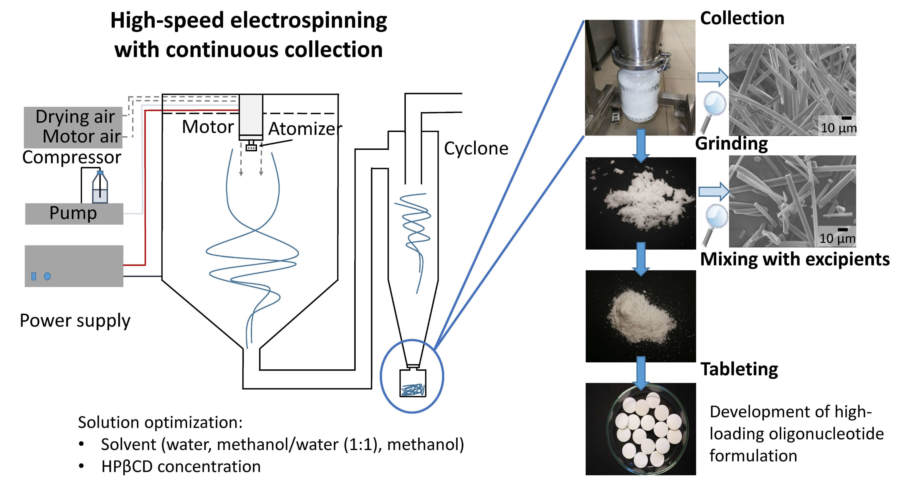
Drugs for oral intake typically come in the form of tablets or capsules. Hirsch et al (2023) used a modified electrospinning setup to produce drug loaded electrospun fibers that can be easily compacted to form a tablet. A multi-orifice rotating spinneret was used for rapid dispensing of the solution. An air dry blower was installed behind the spinneret so that the generated dry air helps to carry the electrospun fibers to a cyclone chamber which compacts the fiber into a glass bottle. This method was used to produce fibers containing antisense oligonucleotides (ASOs) with 2-Hydroxypropyl-beta-cyclodextrin (HPβCD) as the carrier and eventual milling and compression tableting into tablets. An advantage of using electrospinning to produce stable solid forms of sensitive biologicals including oligonucleotides is that it avoided freezing stresses caused by freeze drying. The setup used by Hirsch et al (2023) contained 8 orifices and the rotational speed of the spinneret was 10,000 rpm with an applied voltage of 40 kV. This was able to reach a production rate of ~330 g/h.
Published date: 15 May 2018
Last updated: 09 January 2024
▼ Reference
-
Abdelhakim HE, Coupe A, Tuleu C, Edirisinghe M, Craig DQM. Utilising Co-Axial Electrospinning as a Taste-Masking Technology for Paediatric Drug Delivery. Pharmaceutics. 2021; 13(10):1665.
Open Access
-
Asawahame C, Sutjarittangtham K, Eitssayeam S, Tragoolpua Y, Sirithunyalug B, Sirithunyalug J. Antibacterial Activity and Inhibition of Adherence of Streptococcus mutans by Propolis Electrospun Fibers. AAPS PharmSciTech 2015; 16: 182.
Open Access
-
Colley H E, Said Z, Santocildes-Romero M E.Baker R, D'Apice K, Hansen J, Madsen L S, Thornhill M H, Hatton P V, Murdoch C. Pre-clinical evaluation of novel mucoadhesive bilayer patches for local delivery of clobetasol-17-propionate to the oral mucosa. Biomaterials 2018; 178: 134.
Open Access
-
Ding Y, Dou C, Chang S, Xie Z, Yu DG, Liu Y, Shao J. Core-Shell Eudragit S100 Nanofibers Prepared via Triaxial Electrospinning to Provide a Colon-Targeted Extended Drug Release. Polymers 2020; 12: 2034.
Open Access
-
Dott C, Tyagi C, Tomar L K, Choonara Y E, Kumar P, Toit L C, Pillay V. A Mucoadhesive Electrospun Nanofibrous Matrix for Rapid Oramucosal Drug Delivery. Journal of Nanomaterials 2013; 2013: 924947.
Open Access
Open Access
-
Hirsch E, Nacsa M, Pantea E, Szabó E, Vass P, Domján J, Farkas A, Nyíri Z, Eke Z, Vigh T, et al. Oligonucleotide Formulations Prepared by High-Speed Electrospinning: Maximizing Loading and Exploring Downstream Processability. Pharmaceutics. 2023; 15(3):855.
Open Access
-
Illangakoon U E, Nazir T, Williams G R, Chatterton N P. Mebeverine-Loaded Electrospun Nanofibers: Physicochemical Characterization and Dissolution Studies. Pharm Sci 2014; 103: 283.
-
Li C, Wei Y S, Wen O, Feng K, Zong M H, Wu H. Preparation and characterization of an electrospun colon-specific delivery system for salmon calcitonin. RSC Adv. 2018; 8: 9762.
Open Access
-
Ning T, Zhou Y, Xu H, Guo S, Wang K, Yu D-G. Orodispersible Membranes from a Modified Coaxial Electrospinning for Fast Dissolution of Diclofenac Sodium. Membranes. 2021; 11(11):802.
Open Access
-
Qi M, Li X, Yang Y, Zhou S. Electrospun fibers of acid-labile biodegradable polymers containing ortho ester groups for controlled release of paracetamol. European Journal of Pharmeutics and Biopharmaceutics 2008; 70: 445.
-
Santocildes-Romero M E, Hadley L, Clitherow K H, Hansen J, Murdoch C, Colley H E, Thornhill M H, Hatton P V. Fabrication of Electrospun Mucoadhesive Membranes for Therapeutic Applications in Oral Medicine. ACS Appl. Mater. Interfaces 2017; 9 (13): 11557.
-
Sipos E, Kósa N, Kazsoki A, Szabo Z I, Zelko R. Formulation and Characterization of Aceclofenac-Loaded Nanofiber Based Orally Dissolving Webs. Pharmaceutics 2019; 11(8).
Open Access
-
Topuz F. Rapid Sublingual Delivery of Piroxicam from Electrospun Cyclodextrin Inclusion Complex Nanofibers. ACS Omega 2022; 7: (39), 35083.
Open Access
-
Tyagi C, Tomar L, Choonara Y E, Toit L C, Kumar P, Pillay V. Electrospun Nanofiber Matrix with a Mucoadhesive Backing Film for Oramucosal Drug Delivery. International Journal of Materials, Mechanics and Manufacturing 2014; 2: 81.
-
Vass P, Pantea E, Domokos A.Hirsch E, Domján J, Németh A, Molnár M, Fehér C, Anderson S K, Vigh T, Verreck G, Csontos I, Marosi G, Nagy Z K. Electrospun Solid Formulation of Anaerobic Gut Microbiome Bacteria. AAPS PharmSciTech 2020; 21: 214.
Open Access
-
Vlachou M, Kikionis S, Siamidi A, Kyriakou S, Tsotinis A, Ioannou E, Roussis V. Development and Characterization of Eudragit®-Based Electrospun Nanofibrous Mats and Their Formulation into Nanofiber Tablets for the Modified Release of Furosemide. Pharmaceutics. 2019: 11(9): 480.
Open Access
-
Xiang S, Tang H W, Zhou J, Li X Z. Electrospinning of Hydroxypropyl-beta-cyclodextrin/Polyvinylpyrrolidone Resveratrol-loaded Nanofibers: Preparation and Characterization. Indian J Pharm Sci 2019; 81(4) :618.
Open Access
-
Yang Y, Liu Z P, Yu D G, Wang K, Liu P, Chen X. Colon-specific pulsatile drug release provided by electrospun shellac nanocoating on hydrophilic amorphous composites. International Journal of Nanomedicine 2018; 13: 2395.
Open Access
-
Yu D G, Zhu L M, Branford-White C J, Yang J H, Wang X, Li Y, Qian W. Solid dispersions in the form of electrospun core-sheath nanofibers. International Journal of Nanomedicine 2011; 6: 3271.
Open Access
▲ Close list