Home | About | Contact


Particles Leaching

salt-nanofiber-membrane
Salt particles dispersed in membrane for pore creation

During electrospinning, nanofibers are constantly laid on top of one another and because of their small diameter, most resultant membranes are relatively tightly packed. To introduce large pores within the membrane and to build up its thickness, one simple method is to introduce particles into the membrane during electrospinning. The introduced particles act as temporary scaffolding to increase the spacing between the nanofibers thus giving the membrane volume. The particles may then be removed using water or appropriate solvent leaving behind empty spaces. A suitable material will be salt particles which can easily dissolve in water [Nam et al 2007]. Kim et al drop NaCl salt particulates (size of 100 - 200 microns) onto the deposited fibers at a rate of 10 mg/min/cm2 during electrospinning. The resultant structure with a nanofiber/salt ratios of 10:90 - 20:80 is sufficiently robust to be cut into blocks prior to salt leaching. However, the volume shrunk by 50% after salt removal probably due to partial collapsing of the empty macropores [Kim et al 2008].

wulkersdorfer2010.jpg
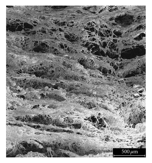
Cross section of electrospun scaffold after leaching of sucrose particles from within [Wulkersdorfer et al. International Journal of Polymer Science, vol. 2010, Article ID 436178, 9 pages, 2010. doi:10.1155/2010/436178. This work is licensed under a Creative Commons Attribution 3.0 Unported License.]

Electrospun scaffolds using sucrose crystals to create the pores was shown to facilitate cell penetration across its thickness. The scaffold was created by alternate electrospinning and deposition of salt crystal at 2 minutes interval. Such short deposition interval allows the crystals to be deposited relatively uniformly throughout the fiber scaffold thickness. However, distinct layers of electrospun membrane was observed when the deposition interval was increased to 10 minutes. Culturing of fibroblasts showed minimal cell penetration in control scaffold and ten minute fiber deposition interval scaffold. When the interval was reduced to two minutes and less, there was greater interconnected pores and good cell penetration was observed [Wulkersdorfer et al 2010].

Published date: 27 August 2012
Last updated: 09 December 2013

Comments
[+]


Google Ad.