Electrospinning is known for its ease of incorporating active substance into fibers and this has led to numerous studies related to this functionality such as release of drugs, fragrance, vitamins and more. Due to favorable cell response to nanofibrous surface, researchers seek to enhance the benefits of electrospun membrane by introducing biomolecules and drugs. Conventional applications such as tissue scaffolds favor controlled and gradual release of biomolecules and drugs which is a challenge given the high surface area to volume of nanofibers. However, there are some applications where rapid release of active substances is required and this makes electrospun nanofibers highly relevant and attractive. Due to the high surface area of electrospun nanofibers, its membrane release rate is expected to be faster than cast film. Seif et al (2016) compared the release rate of lysozyme from electrospun poly(vinyl alcohol) (PVA) fibers and PVA cast film in PBS using fiber-optics based dissolution testing system for in situ monitoring of drug release. Their study showed that 90% of the drug was released from electrospun fibers membrane within 6 minutes while it takes about 15 minutes from cast film to release the same amount.
An advantage of drug encapsulation in electrospun fibers is that the spinning process reduces drug crystallization. Drug crystallization has been said to make the drug delivery system unstable and causes a drop in the drug loading efficiency and unpredictable release behavior [Kwak et al 2017]. Using water soluble fish gelatin (FG), Kwak et al (2017) demonstrated the lack of crystallization and the rapid release of caffeine in electrospun FG. For solvent cast FG film, with only 0.8 w/v% of caffeine added, spherical crystalline nucleus can be seen. Electrospun fibers in contrast can take up to 2% loading in FG without any disruption to the smooth fiber morphology. Repeated folding of the electrospun caffeine loaded FG membrane without any cracks but solvent cast FG film cracks after only folding 5 times. Total disintegration in water only took 1.5 s in electrospun FG/caffeine membrane but took 40 s in solvent cast film form. On wetted sponge, total disintegration of electrospun FG/caffeine membrane took 5 s but solvent cast film maintained in gel state until 3 minutes due to inadequate water absorption for total disintegration.
Oral drug delivery is an area where rapid release of active pharmaceutical ingredients is required. There are several advantages and recommendations for oral drug delivery,
- Accurate dosage
- Instant drug release (eg. pain relieve)
- Suitable for patient with swallowing difficulty
- Buccal and oral track treatment
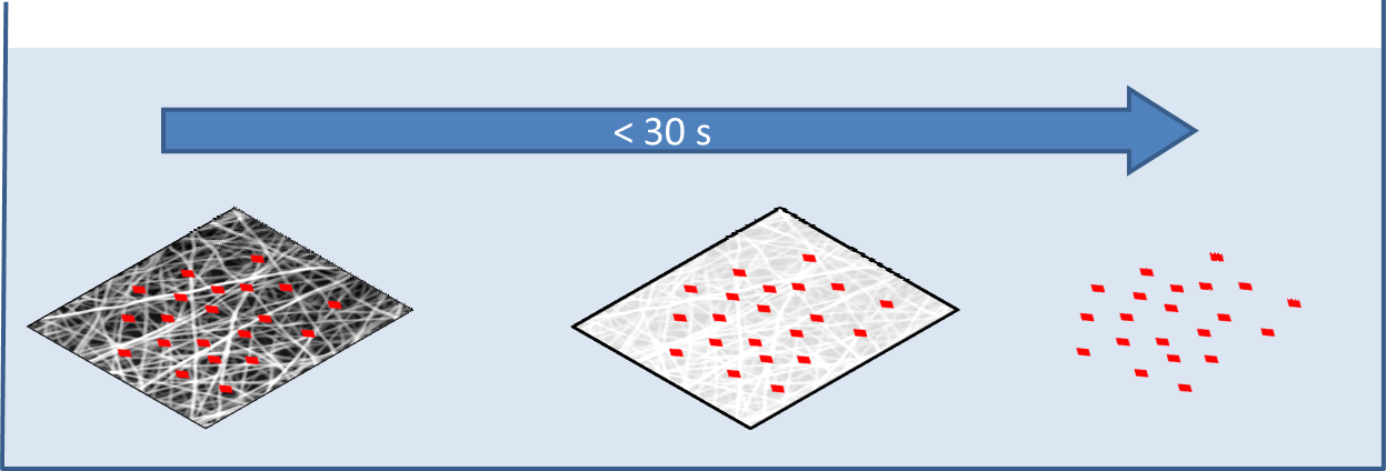
Liquid and loose powders are generally poor for delivery of accurate dosage. Orally disintegrating tablet formulation may be common but the level of compaction is a compromise between having a sufficient holding strength (to prevent disintegration) and rapid disintegration in oral environment. Oral delivery in the form of film offers advantages such as having a larger surface area and mechanically flexible and is currently available in the market. Electrospun film with its higher surface area is potentially better than smooth film for rapid drug release. Nagy et al (2010) compared the release of Donepezil HCI in loaded electrospun polyvinyl alcohol (PVA) film, smooth PVA film and commercial Donepezil HCI tablet. Full release of Donepezil HCI from PVA film took less than 10 s while it takes about 30 min for smooth film and tablet.
For mucoadhesive drug delivery where topical application is preferred, ultrafast release of the drug alone is insufficient as the patient may ingest most of them. To facilitate optimal retention of the drug in the area of application, a less soluble backing material may be used [Dott et al 2013; Tyagi et al 2014]. Dott et al (2013) electrospun polyvinyl alcohol (PVA) loaded with citric acid, diphenhydramine and glycerol on a film cast from PVA and hydroxypropylmethylcellulose (HPMC). While the electrospun film alone takes less than 4 s for complete disintegration, cast film with electrospun fiber took about 13 s. Cast film alone with the same components and composition took about the same time for complete disintegration but its mechanical strength is weaker than cast film with electrospun coating. To impart different functionality to the fiber, a core-sheath fiber structure may be used. Although core-sheath fiber structure is more commonly used to control the release rate of drugs, a highly water soluble sheath material will also allow rapid release of the drugs located in the core of the fiber. Yu et al (2011) constructed a core-sheath fiber with the sheath comprising of PVP and sucralose as sweetener and core material comprising of PVP and a drug, acyclovir. In vitro results showed that all acyclovir were released in less than a minute but the release rate of acyclovir powder is much longer with less than fifty percent released after an hour. Slow release rate of acyclovir powder probably due to the size of the particles which is about 100 µm. Asawahame et al (2015) investigated the potential use of water-soluble polyvinylpyrrolidone nanofibers loaded with propolis (PVP-propolis) as antibacterial agent in the mouth. Pure PVP-propolis nanofibers were found to be completely wetted in 45s while the addition of Tween 80 and small amount of flavouring agents reduced the wetting time to less than 2s. When tested against Streptococcus mutans (S. mutans), PVP-propolis nanofibers containing 0.6 MIC (minimum inhibitory concentration, 1.172 mg/mL) were effective in decreasing the adherence of the bacteria to the glass surface. The addition of Tween 80 and flavouring agents does not show significant difference in the inhibition of S. mutans adherence to glass compared to fibers without.
Another consideration is the interaction between the drug and the polymer. Yu et al (2009) found that when ibuprofen was blended with polyvinylpyrrolidone (PVP) solution and electrospun to form fibers, hydrogen bond is formed between ibuprofen and PVP when ibuprofen was at low concentration (1:4 ratio of ibuprofen/PVP). When the concentration of ibuprofen was increased, ibuprofen starts to form aggregates within the fiber. Upon contact with water, although disintegration of the membrane is rapid (less than 10 s) and independent of the amount of drug loaded, the dissolution of ibuprofen differs significantly. At lower drug concentration, drug dispersion into water is easier as the drug was released as molecules instead of as aggregates which were the case for higher loading.
Since water soluble polymers are often used in rapid drug delivery, this may limit the amount of water-insoluble active substances that can be loaded into the fiber. A possible option is to use cyclodextrin which can form complexes with hydrophobic compounds and yet retain high solubility in water. Manasco et al (2014) was able to load poorly water soluble ketoprofen into PVA solution with hydroxypropyl-β-cyclodextrin (HPβCD). A high HPβCD content can be blended with PVA to give a ratio of 90:10. This allows ketoprofen loading of about 7% into the electrospun fibers and a dissolution time of 10 s was recorded. Similarly, Borbas used sulfobuthyletherβ-cyclodextrin (SBEβCD) for encapsulation of aripiprazole (ARP) in the presence of citric acid in poly(ethylene oxide) (PEO) solution for electrospinning into fibers. The combination was optimized such that the lowest possible PEO concentration for fiber production was used as PEO may retard the dissolution process. Comparing the dissolution time taken for the ARP loaded PEO, pure ARP and mixture of ARP and SBEβCD, it takes only 3 minutes for complete dissolution of ARP loaded PEO electrospun fibers. The mixture only released 20% of ARP for the same duration while dissolution of pure ARP was less than 3% after 60 minutes. Faster release rate of ARP in electrospun fibers is because the drug remains in an amorphous state instead of thermodynamically stable crystalline form. ARP encapsulated in electrospun PEO fibers was found to remain in amorphous state even after 3 months.
Topuz et al (2022) was able to electrospin pure HPβCD for loading of Piroxicam (Px), a nonsteroidal anti-inflammatory drug. The solution was prepared in water mixing Px and HPβCD for a day hence there is no use of potentially toxic solvents. Smooth fibers of diameters 500 nm or less were produced from electrospinning the solution with higher loading of Px leading to smaller fiber diameters. Px in the HPβCD fibers were found to form inclusion complexes and the initial highly crystalline Px became amorphous as demonstrated by the absence of sharp peaks in XRD patterns. The resultant Px embedded nanofibrous membrane may be used for rapid sublingual delivery of Px. When placed in artificial saliva, the dissolution of the membrane was completed within a second and about 90% of Px was released within a minute and close to 100% of the Px was released after 10 min. Px in the fibers were found to be stable after six months of storage which can be attributed to the complexation between the CD and Px.
Another option is to use core-shell electrospinning where the shell material is water soluble and the core material containing water insoluble drug. Li et al (2013) used this technique to incorporate quercetin into the core of polyvinylpyrrolidone fibers with loading of up to 16.5%. Upon contact with saline water, the nanofibrous film quickly disintegrates and release quercetin into the solution.
Water soluble polymers are often used for drug encapsulation due to its rapid solubility in water. However, it is also important to consider the pH of the environment which it is to be used in. Jain et al (2014) showed that the drug release rate of polyvinylpyrrolidone (PVP) nanofibers varied with pH. In vitro release of Glibenclamide (GLB) from PVP nanofibers was found to be 82%, 95% and 24% at pH of 7.8, 6.4 and 4 respectively at 5 minutes. For oral application, fluctuation of the saliva pH [Aframian et al 2006] may need to be taken into account to have a complete understanding of the release rate.
Apart from oral drug delivery, ultrafast release of active substance may also be used in the prevention of human immunodeficiency virus (HIV) as an intra-vaginal drug delivery vehicle. Huang et al (2012) electrospun cellulose acetate phthalate (CAP) fibers loaded with anti-viral substances. CAP was selected as it is stable in low pH which is the condition found in healthy vaginal environment but readily soluble when the pH increases to 5. As low as 20% semen in simulated vaginal fluid was found to sufficiently increase the pH such that significant amount of CAP dissolves. With 30% semen in simulated vaginal fluid, the CAP dissolves in 18 s. Loading of the fiber with just 0.5 µg Viread/ml was able to achieve complete neutralization of HIV-1 infection.
Electrospun nanofibers are a good carrier for quick release of enzyme for applications such as microfluidic detection chip. High surface area of the nanofibers allows rapid dissolution in appropriate water or solvents. Dai et al (2012) used water soluble electrospun polyvinyl pyrrolidone nanofibers as carrier for horseradish peroxidises (HRP). Placed in a microfluidic chip, PVP nanofibers loaded with HRP readily dissolve to release HRP when the aqueous sample passed through it.
Electrospinning may also be used as a drying technology for encapsulating biological agents. Conventional technology such as freeze drying may affect the viability of proteins and other biological agents. While many of these biological agents are administered in a solution form, having them dried during storage can improve their shelf-life. Biological agents may be dried for storage but it is important that its viability or the efficacy does not deteriorate significantly during storage. Hirsch et al (2021) tested several excipients loaded into electrospun fibers on their ability to maintain viability of encapsulated microbes. Excipients may afford protection due to their amorphous state by restricting molecular movement in their glassy matrix which may damage the microbe. Other excipients may be prebiotics which may help the maintenance of the microbes. Hirsch et al (2021) used Lactobacillus paracasei blended in polyvinyl alcohol (PVA)/polyethylene oxide(PEO) solution and stabilizing excipient (glucose, lactose, mannitol, saccharose, trehalose, inulin) and electrospinning. Saccharose, trehalose and skim milk were found to be the most effective excipients and showed similar or better results compared to other drying techniques. L. paracasei dried using electrospinning and skim milk as excipient has a survival rate of 85% compared to just 32% using spray drying using skim milk. Lower temperature was also found to enhance storage survival rate. The viability of the bacteria drops to zero in 7 to 120 days when stored at 25 °C. However, the bacteria remains viable for 1 year of storage at 7 °C and -20 °C after which storage at -20 °C showed better survival rate. The lesser viability at higher storage temperature has been attributed to the movement of molecules at higher temperature which may damage cell walls. Dissolution of the PVA/PEO fibers containing the excipients and microbes were rapid with complete dissolution in 1s. Gelation of PVA was not seen and this has been attributed to high loading of water soluble excipients.
High surface area of electrospun fibers and using water soluble polymers as encapsulation agents meant that the loaded fibers can be dissolved quickly for reconstituting the biological agent. Domján et al (2020) electrospun water soluble 2-hydroxypropyl-β-cyclodextrin (HP-β-CD) fibers loaded with infliximab antibody. Infliximab remained viable with no sign of degradation after electrospinning with HP-β-CD. The electrospun HP-β-CD fibers containing infliximab were able to completely dissolve in water after 120s without any vigorous mixing. This makes it convenient and fast to reconstitute infliximab prior to usage.
Dowlath et al (2021) demonstrated the potential use of electrospinning to encapsulate virus-like particle (VLP) vaccine. This VLP is derived from rabbit hemorrhagic disease virus modified to carry the MHC-I gp100 tumor-associated antigen epitope. The VLP was blended into a polyvinylpyrrolidone (PVP) solution followed by electrospinning into fibers. The electrospinning process dries the VLP while the polymer matrix protects the VLP from environmental damage. Prior to vaccination, the VLP loaded nanofiber samples were dissolved in PBS and added with CpG oligonucleotide as an adjuvant. In vivo tests were carried out on mice by subcutaneous administration to the left flank. VLP released by electrospun nanofibers was shown to induce comparable antibody titers to that of the VLP delivered in PBS. This shows that electrospinning can be used to process VLP into a dry formulation nanofiber for reconstituting prior to delivery and maintaining its immunogenicity.
Published date: 26 August 2014
Last updated: 05 Sepember 2023
▼ Reference
-
Aframian D J, Davidowitz T, Benoliel R. The distribution of oral mucosal pH values in healthy saliva secretors. Oral Dis. 2006; 12: 420.
-
Asawahame C, Sutjarittangtham K, Eitssayeam S, Tragoolpua Y, Sirithunyalug B, Sirithunyalug J. Antibacterial Activity and Inhibition of Adherence of Streptococcus mutans by Propolis Electrospun Fibers. AAPS PharmSciTech 2015; 16: 182.
Open Access
-
Borbas E, Balogh A, Bocz K, Muller J, Kiserdei E, Vigh T, Sinko B, Marosi A, Halaz A, Dohanyos Z, Szente L, Balogh G T, Nagy Z K. In vitro dissolution-permeation evaluation of an electrospun cyclodextrin-based formulation of aripiprazole using µFluxTM. International Journal of Pharmaceutics 2015; 491: 180.
-
Dai M, Jin S, Nugen S R. Water-Soluble Electrospun Nanofibers as a Method for On-Chip Reagent Storage. Biosensors 2012; 2: 388.
Open Access
-
Domján J, Vass P, Hirsch E, Szabó E, Pantea E, Andersen S K, Vigh T, Verreck G, Marosi G, K. Nagy Z K. Monoclonal antibody formulation manufactured by high-speed electrospinning. International Journal of Pharmaceutics 2020; 591: 120042.
Open Access
-
Dott C, Tyagi C, Tomar L K, Choonara Y E, Kumar P, Toit L C, Pillay V. A Mucoadhesive Electrospun Nanofibrous Matrix for Rapid Oramucosal Drug Delivery. Journal of Nanomaterials 2013; 2013: 924947.
Open Access
-
Dowlath S, Campbell K, Al-Barwani F, Young VL, Young SL, Walker GF, Ward VK. Dry Formulation of Virus-Like Particles in Electrospun Nanofibers. Vaccines. 2021; 9(3):213.
Open Access
-
Hirsch E, Pantea E, Vass P, Domján J, Molnár M, Suhajda A, Andersen S K, Vigh T, Verreck G, Marosi G J, Nagy Z K. Probiotic bacteria stabilized in orally dissolving nanofibers prepared by high-speed electrospinning. Food and Bioproducts Processing 2021; 128: 84.
Open Access
-
Huang C, Soenen S J, Gulck E, Vanham G, Rejman J, Calenbergh S, Vervaet C, Coenye T, Verstraelen H, Temmerman M, Demeester J, Smedt S C. Electrospun cellulose acetate phthalate fibers for semen induced anti-HIV vaginal drug delivery. Biomaterials 2012; 33: 962.
-
Jain K, Awasthi S, Kumar P, Somashekariah B V, Phani A R. Formulation and Pharmacokinetic Studies of Rapidly Dissolving Nanofibers. Middle-East J Sci Res 2014; 22: 1176.
Open Access
-
Kwak H W, Woo H, Kim I C, Lee K H. Fish gelatin nanofibers prevent drug crystallization and enable ultrafast delivery. RSC Adv., 2017, 7, 40411.
Open Access
-
Li X Y, Li Y C, Yu D G, Liao Y Z, Wang X. Fast Disintegrating Quercetin-Loaded Drug Delivery Systems Fabricated Using Coaxial Electrospinning. Int. J. Mol. Sci. 2013; 14: 21647.
Open Access
-
Manasco J L, Tang C, Burns N A, Saquing C D, Khan S A. Rapidly dissolving poly(vinyl alcohol)/cyclodextrin electrospun nanofibrous membranes. RSC Adv. 2014; 4: 13274.
-
Nagy Z K, Nyul K, Wagner I, Molnar K, Marosi G. Electrospun water soluble polymer mat for ultrafast release of Donepezil HCl. eXPRESS Polymer Letters 2010; 4: 763.
Open Access
-
Seif S, Graef F, Gordon S, Windberg M. Monitoring Drug Release from Electrospun Fibers Using an In Situ Fiber-Optic System. Dissolution Technologies 2016; 23: 6.
Open Access
-
Topuz F. Rapid Sublingual Delivery of Piroxicam from Electrospun Cyclodextrin Inclusion Complex Nanofibers. ACS Omega 2022; 7: (39), 35083.
Open Access
-
Tyagi C, Tomar L, Choonara Y E, Toit L C, Kumar P, Pillay V. Electrospun Nanofiber Matrix with a Mucoadhesive Backing Film for Oramucosal Drug Delivery. International Journal of Materials, Mechanics and Manufacturing 2014; 2: 81.
-
Yu D G, Shen X X, Branford-White C, White K, Zhu L M, Bligh S W A. Oral fast-dissolving drug delivery membranes prepared from electrospun polyvinylpyrrolidone ultrafine fibers. Nanotechnology 2009; 20: 055104.
-
Yu D G, Zhu L M, Branford-White C J, Yang J H, Wang X, Li Y, Qian W. Solid dispersions in the form of electrospun core-sheath nanofibers. International Journal of Nanomedicine 2011; 6: 3271.
Open Access
▲ Close list