
▼ Reference
- Castro-Mayorga J L, Fabra M J, Cabedo L, Lagaron J M. On the Use of the Electrospinning Coating Technique to Produce Antimicrobial Polyhydroxyalkanoate Materials Containing In Situ-Stabilized Silver Nanoparticles. Nanomaterials 2017; 7(1): 4 Open Access
- Chaudhary A, Gupta A, Mathur R B, Dhakate S R. Effective antimicrobial filter from electrospun polyacrylonitrile-silver composite nanofibers membrane for conducive environment. Adv. Mat. Lett. 2014; 5: 562. Open Access
- Jin S, LI J, Wang J, Jiang J, Zhou Y, Li Y, Yang F. Electrospun silver ion-loaded calcium phosphate/chitosan antibacterial composite fibrous membranes for guided bone regeneration. International Journal of Nanomedicine 2018; 13: 4591. Open Access
- Liang S, Zhang G, Min J, Ding J, Jiang X. Synthesis and Antibacterial Testing of Silver/Poly (Ether Amide) Composite Nanofibers with Ultralow Silver Content. Journal of Nanomaterials 2014; 2014: 684251. Open Access
- Lin S, Wang R, Yi Y, Wang Z, Hao L, Wu J, Hu G, He H. Facile and green fabrication of electrospun poly(vinyl alcohol) nanofibrous mats doped with narrowly dispersed silver nanoparticles. International Journal of Nanomedicine 2014; 9: 3937. Open Access
- Lok C N, Ho C M, Chen R, He Q Y, Yu W Y, Sun H, Tam P W H, Chiu J F, Che C M. Silver nanoparticles: partial oxidation and antibacterial activities. Journal of Biological Inorganic Chemistry 2007; 12: 527.
- Nuge T, Tshai K Y, Lim S S, Nordin N, Hoque M E. Preparation and characterization of Cu-, Fe-, Ag-, Zn- and Ni- doped gelatin nanofibers for possible applications in antibacterial nanomedicine. Journal of Engineering Science and Technology 5th EURECA 2015 Special Issue March 2017; 3: 68. Open Access
- Preethi G U, Unnikrishnan B S, Joseph M M, Shiji R, Sreelekha T T. Biogenic silver nanoparticles embedded polyvinyl alcohol nanofibrous scaffolds avert tumour and bacterial growth. Research Communications. 2019; 116: 1735.
- Son W K, Youk J H, Park W H. Antimicrobial cellulose acetate nanofibers containing silver nanoparticles. Carbohydrate Polymers 2006; 65: 430.
- Rujitanaroj P O, Pimpha N, Supaphol P. Preparation, Characterization, and Antibacterial Properties of Electrospun Polyacrylonitrile Fibrous Membranes Containing Silver Nanoparticles. J Appl Polym Sci 2010; 116: 1967.
- Rujitanaroj P O, Pimpha N, Supaphol P. Wound-dressing materials with antibacterial activity from electrospun gelatin fiber mats containing silver nanoparticles. Polymer 2008; 49: 4723.
- Yuan J, Geng J, Xing Z, Shen J, Kang I K, Byun H. Electrospinning of Antibacterial Poly(vinylidene fluoride) Nanofibers Containing Silver Nanoparticles. J Appl Polym Sci 2010; 116: 668.
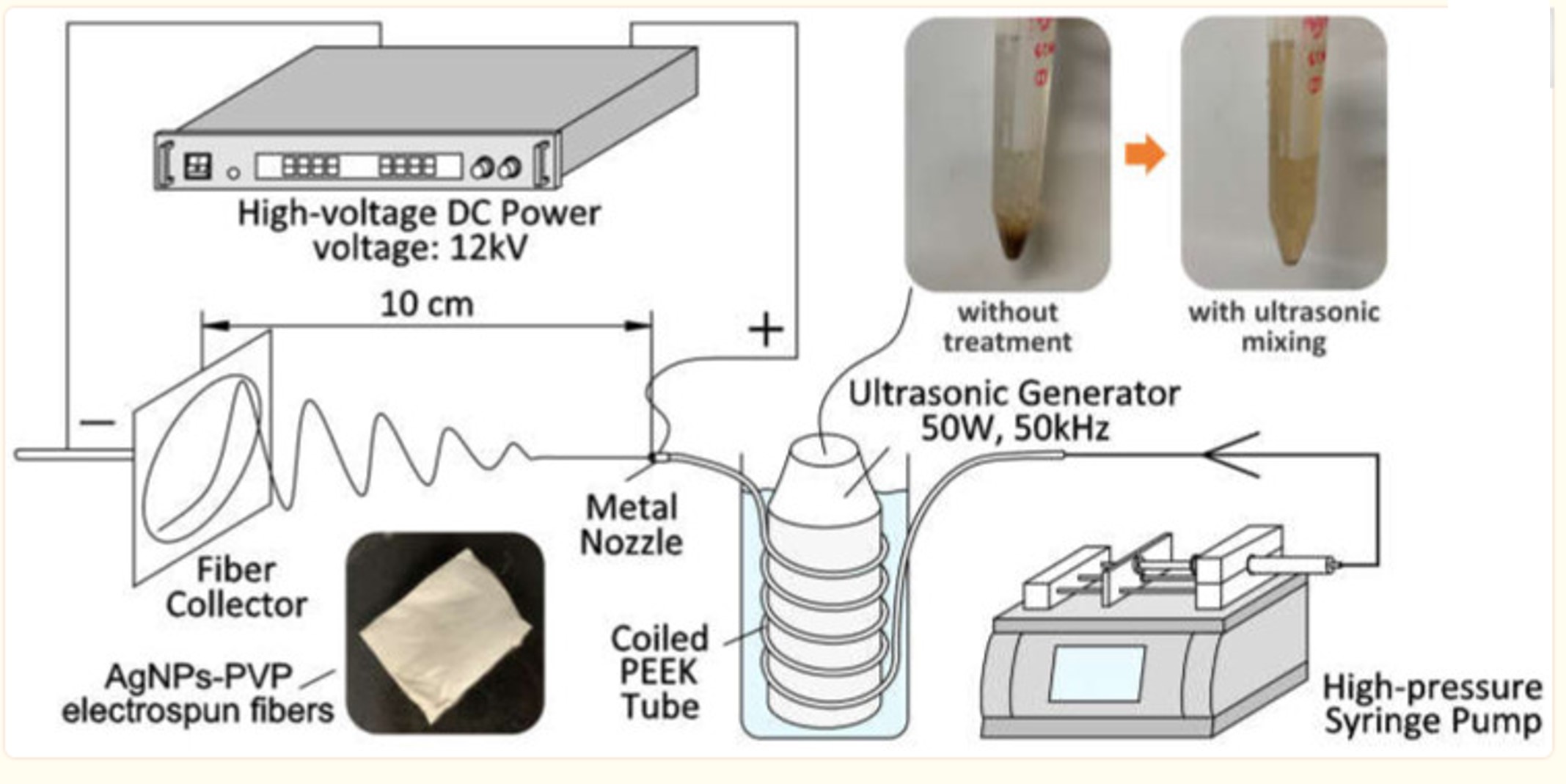
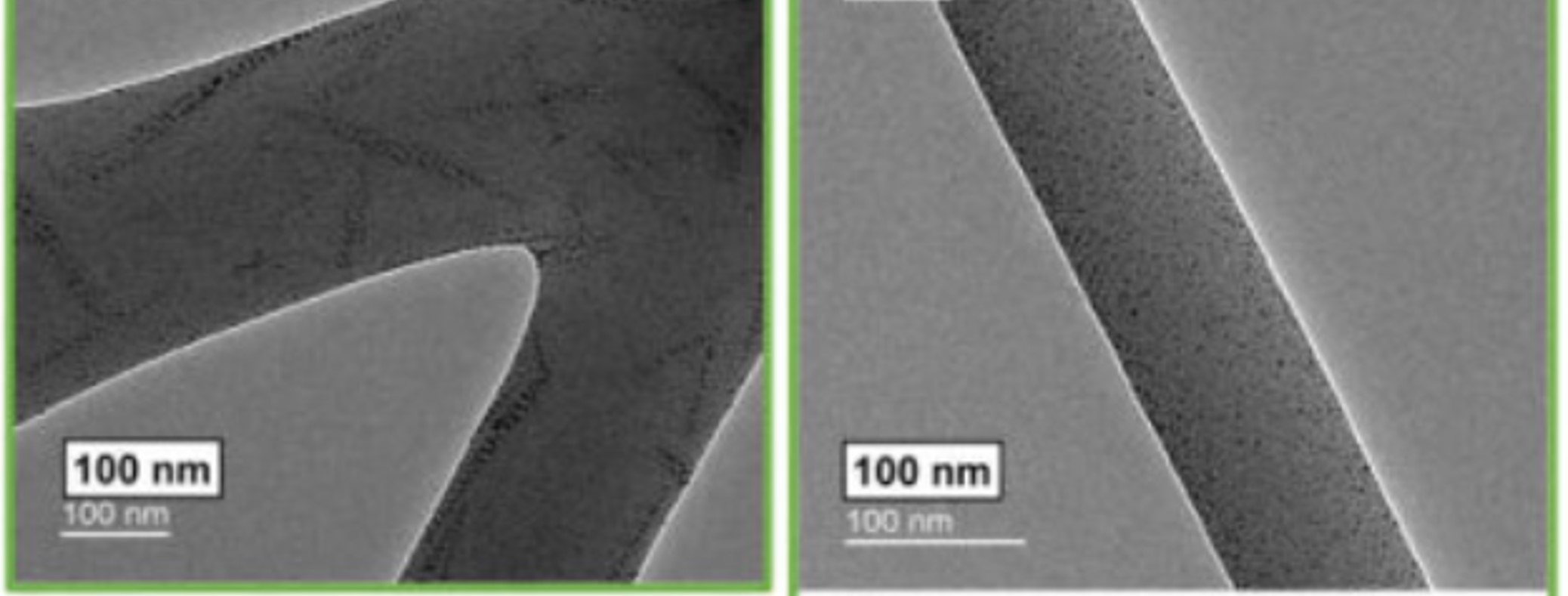
- Zhu L, Zhu W, Hu X, Lin Y, Machmudah S, Wahyudiono, Kanda H, Goto M. PVP/Highly Dispersed AgNPs Nanofibers Using Ultrasonic-Assisted Electrospinning. Polymers (Basel). 2022; 14(3): 599 Open Access
▼ Credit and Acknowledgement
Author
Wee-Eong TEO View profile
Email: weeeong@yahoo.com
