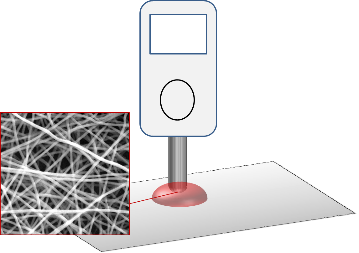
Glucose sensor is often based on incorporating glucose oxidase into a device and measuring the electrical current. Electrospun fibers may be used for different function in this setup. The range of materials available for electrospinning makes it feasible to incorporate glucose oxidase on the nanofibers. For implantable glucose sensors, electrospun fibers may also be used as a carrier for drugs to promote integration with the sensing device.
Various materials have been electrospun for incorporation of glucose oxidase onto it. In most cases, electrospun nanofibers containing glucose oxidase are coated on a conducting electrode to determine the current output. The simplest method of having glucose oxidase in the nanofibers is by mixing glucose oxidase into the solution and electrospinning. This has been shown to be adequate in detecting current changes when the electrospun coated electrode is immersed into glucose solution. Wu et al (2014) showed that by adding graphene to the glucose oxidase/polyvinyl alcohol (PVA) solution, the sensitivity of the glucose sensor is improved. Presence of graphene was found to stabilize enzyme's conformation and facilitate the catalytic reaction. However when the graphene content exceeds 20 ppm, performance of the glucose sensor drops [Wu et al 2014]. It is not immediately clear why this is so. While incorporation of glucose oxidase through blending is the simplest method, there is a risk that glucose oxidase may leach out of the polymer matrix. The sensitivity of the nanofiber sensor may be reduced as the glucose oxidases are encapsulated within the polymer matrix.
Having glucose oxidase bonded on the surface of the nanofibrous sensor is likely to improve sensor sensitivity. Immobilization of glucose oxidase may be achieved by electrostatic interaction between negative charges in the enzyme with a positively charged material. Manesh et al (2008) used positively charged multi-walled carbon nanotube (MWCNT) wrapped in a cationic polymer (poly(diallyldimethylammonium chloride) (PDDA)) to immobilize glucose oxidase on the surface of polymethylmethacrylate (PMMA) nanofiber. The process involved electrospinning a blend of MWCNT/PDDA/PMMA onto an ITO-coated glass plate and incubating the assembly in glucose oxidase solution. To improve selectivity against interfering species such as ascorbic acid and uric acid, a thin layer of nafion was coated. Negatively charged nafion is able to electrostatically repel negatively charged ascorbic acid and uric acid [Manesh et al 2008]. Hu et al (2019) constructed a glucose biosensor based on TiO2 nanofibers. TiO2 nanofibers (p-TDNF) were synthesized by electrospinning a tetrabutyl titanate (TBT), polyvinylpyrrolidone precursor solution and calcined at 500°C. An electron beam treatment on p-TDNF introduces surface defect which makes the TiO2 nanofibers more reactive (mTDNF). The electron beam irradiation also form Ti3+ on the nanofibers and together with the surface defects, lowers the band gap width of mTDNF to 2.846 eV, a reduction of 0.36 eV from pTDNF. This reduces the transmission barrier and increases electron diffusion path. Surface defects and Ti3+ also helps to inhibit recombination of electron-hole pairs and this enhances chemical activity. When tested for use as glucose biosensor, glucose oxidase (GOD)/m-TDNF/Nafion/ glassy carbon electrode (GCE) assembly showed anode and cathode response currents that were 1.6 times higher than that of GOD/p-TDNF/Nafion/GCE. This has been attributed to the stronger electrostatic interaction between GOD and mTDNF. The (GOD)/m-TDNF/Nafion/ glassy carbon electrode (GCE) biosensor also showed a higher sensitivity of 12.5 µA.mM-1cm-2, a low limit of detection of 0.9 µM and fast current response of less than 3s.
For better sensitivity the material should quickly transfer the electron generated to the reader. Huang (2009) constructed Mn2O2-Ag nanofibers for immobilization of the glucose oxidase. The device showed excellent selectivity with negligible response to presence of uric acid (UA) and ascorbic acid (A A) at their physiological concentration level.
| Electrospun Material |
Sensitivity |
Reference |
| Mn2O2-Ag |
40.6µA.cm-2.mM-1 |
Huang et al 2009 |
| ZnO-CuO nanocomposite |
3066.4µA.cm-2.mM-1 |
Zhou et al 2014 |
| Polyvinyl alcohol (PVA)/glucose oxidase/graphene |
38.7µA.mM-1 |
Wu et al 2014 |
Non-enzymatic approach has also been investigated for glucose detection using electrospun nanofibers. Liu et al (2009) constructed a renewable electrospun Ni nanoparticle-loaded carbon nanofiber paste electrode. Detection is through measuring the electrocatalytic activity of the Ni nanoparticles towards glucose oxidation.
Li et al ((2023) constructed Mn3O4/NiO nanoparticles decorated on carbon nanofibers (CNF) as a sensor for glucose detection. The solution for electrospinning was a blend of polyacrylonitrile (PAN) with Ni salt and Mn salt as precursor materials. Heat treatment was carried out for carbonization of PAN to form carbon nanofiber and the salts to Mn3O4 and NiO nanoparticles. Comparing the electrochemical performance of Mn3O4/NiO/CNF with Mn3O4/CNF and NiO/CNF, Mn3O4/NiO/CNF showed the the fastest charge transfer capability hence demonstrating the synergistic interaction between Mn and Ni in the oxidation of glucose. The Mn3O4/NiO/CNF sensor was able to maintain good selectivity with negligible current response with 5% interference agent added. Long term stability of the sensor was demonstrated by a reduction of less than 5% of the initial current after 30 days storage in room temperature.
Zhou et al (2014) constructed a three-dimensional network of ZnO-CuO nanocomposites by electrospinning for the detection of glucose. This works by a Cu(II)/Cu(III) redox couple,
CuO + OH- - e- → CuO(OH)
Presence of glucose gives Cu(III) ions an electron which is released to the electrode where it will be registered as a rise in the peak current. The ZnO-CuO nanofibrous nanocomposites demonstrated good sensitivity and selectivity against common interfering species such as UA, AAm, Mg2+ and others [Zhou et al 2014].
Protection of implantable glucose sensors from inflammatory response and bacterial infections is essential for its long term reliability. Nitric oxide (NO) may be used to mitigate foreign body response and to foster integration with the host tissues. Koh et al (2011) used electrospun polyurethane as a carrier for the release of NO to protect the platinum electrodes pre-coated with glucose oxidase. An advantage of electrospun fibers as carrier versus cast film carrier is the highly porous nature of electrospun fiber membrane which allows better permeability of O2 and H2O2 as given by its sensitivity of 150 nA/M and response time of less than a minute.
Glucose sensors may also be constructed in the form of wearable devices. For this, the sensor needs to be able to detect small amounts of glucose. Kim et al (2020) showed that glucose oxidase (GOx) blended into poly(vinyl alcohol) (PVA) aqueous solution followed by the cross linking agent 1,2,3,4-butanetetracarboxylic acid (BTCA), when electrospun produced nanofibers with very low enzyme activity. However, when GOx was allowed to form complexes with β-cyclodextrin (β-CD) prior to blending with PVA and BTCA for electrospinning and post spinning heat treatment, the enzymatic activity improved significantly to 59.3%. The enzyme activity was further increased to 76.3% with the addition of Au nanoparticles (AuNPs) into the PVA/BTCA/β-CD/AuNPs/GOx hydrogel, possibly due to better conductivity. The constructed sensor made from PVA/BTCA/β-CD/GOx hydrogel showed a linear range for the glucose concentration from 0.1 mM to 0.5 mM with a sensitivity of 47.2 µA mM-1 and a detection limit of 0.01 mM.
Published date: 12 May 2015
Last updated: 05 December 2023
▼ Reference
-
Hu Z, Rong J, Zhan Z, Yu X. Biosensor based on TiO2 Nanofibers Prepared Using High Energy Electron Beam Treatment for Rapid and Efficient Glucose Determination. Int. J. Electrochem. Sci. 2019; 14: 10352.
Open Access
-
Huang S. Glucose Biosensor Using Electrospun Mn2O3-Ag Nanofibers. Master's Theses. 2011 Paper 147 Hunan Normal University.
Open Access
-
Kim G J, Kim K O. Novel glucose-responsive of the transparent nanofiber hydrogel patches as a wearable biosensor via electrospinning. Sci Rep 2020; 10: 18858.
Open Access
-
Koh A, Riccio D A, Sun B, Carpenter A W, Nicholas S P, Schoenfisch M H. Fabrication of nitric oxide-releasing polyurethane glucose sensor membranes. Biosens Bioelectron 2011; 28: 17.
Open Access
http://www.ncbi.nlm.nih.gov/pmc/articles/PMC3186917/
-
Li M, Dong J, Deng D, Ouyang X, Yan X, Liu S, Luo L. Mn3O4/NiO Nanoparticles Decorated on Carbon Nanofibers as an Enzyme-Free Electrochemical Sensor for Glucose Detection. Biosensors. 2023; 13(2):264.
Open Access
-
Liu Y, Teng H, Hou H, You T. Nonenzymatic glucose sensor based on renewable electrospun Ni nanoparticle-loaded carbon nanofiber paste electrode. Biosensors and Bioelectronics 2009; 24: 3329.
-
Manesh K M, Kim H T, Santhosh P, Gopalan A I, Lee K P. A novel glucose biosensor based on immobilization of glucose oxidase into multiwall carbon nanotubes-polyelectrolyte-loaded electrospun nanofibrous membrane. Biosensors and Bioelectronics 2008; 23: 771.
-
Wu C M, Yu S A, Lin S L. Graphene modified electrospun poly(vinyl alcohol) nanofibrous membranes for glucose oxidase immobilization. eXPRESS Polymer Letters 2014; 8: 565.
Open Access
-
Zhou C, Xu L, Song J, Xing R, Xu S, Liu D, Song H. Ultrasensitive non-enzymatic glucose sensor based on three-dimensional network of ZnO-CuO hierarchical nanocomposites by electrospinning. Scientific Reports 2014; 4: 7382.
Open Access
▲ Close list
 ElectrospinTech
