Home | About | Contact


Electrospun fibers as implant interface layer

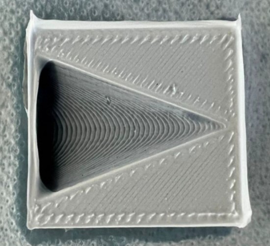
Electrospun nanofibers are known to facilitate cell adhesion and proliferation in numerous in vitro tests. In vivo studies has also shown low inflammatory reactions. Due to the varied demands of implantables, it is not possible to use electrospun nanofibers in all situations. However, the advantages of electrospun fibers may be employed to facilitate integration between implants and surrounding host tissues. Electrospun membrane may be used as a barrier against adhesion between tissues and organs during surgery. They may also be used to reduce infection through incorporation of drugs and to reduce immune response on the implant. With electrospinning it is important that the collecting surface exhibits a minimum level of conduction for the charged fibers to deposit on it. Polylactic acid (PLA) is a common biodegradable material that can be used for 3D printing into implantable scaffolds. However, pure PLA is a very poor electrical conductor and it is not possible to use electrospinning to directly deposit fibers on it. Conductive PLA has been developed for use in 3D printing and electrospinning has been shown to be able to deposit nanofibers on its printed scaffolds [Bauer et al 2022]. One challenge of electrospinning fibers on implant is that fibers may not deposit in cavities or concave part of an undulating surface. Electrospun fibers deposit at the point where the electric field lines are the strongest. With conductive PLA material, fibers can still deposit on the lower concave part of the implant as the conductivity of the material is not so strong that the contrast in the potential field between the different elevations causes the fibers to deposit across the cavity instead of in it [Buer et al 2022]. Similarly, a lower applied voltage was also found to allow fiber deposition in lower areas [Buer et al 2022].


PAN electrospun on a funnel with a length of 40 mm and width of 31 mm, printed with Conductive PLA [Bauer et al 2022].


Facilitate Integration

Metal is a commonly used material as collector for electrospun fibers, thus it is relatively straight forward to coat a metal implant with electrospun nanofibers. This has been demonstrated on titanium implant [Ravichandran 2009] and electrospun fibers have also been deposited on magnesium alloy sheet [Soujanya et al 2014]. To enhance integration, mineralization has been carried out on the nanofibers to form polymer-hydroxyapatite composite. Collagen is often used as one of the components in the nanofibers to enhance mineralization [Iafisco et al 2012, Ravichandran 2009]. Ravichandran (2009) showed that adhesion of mesenchymal stem cells (MSC) on titanium plate and alloy is significantly enhanced with nanofiber coating even with man-made polymer such as poly(lactic acid)-co-poly(glycolic acid) (PLGA). In an alternative application, Liao et al (2017) used melt electrospinning to coat polycaprolactone (PCL) fibers on a silicone base layer. This bilayer construct is designed to create a seal on the interface in a novel suture-less inflow cannula. The presence of electrospun PCL fibers is to facilitate tissue integration. Culture with human foreskin fibroblasts showed good adhesion to the electrospun layer in contrast to low cell adhesion on the flat silicone sheet. Proliferation of the cells on bilayered scaffold was also significantly better than silicone sheet over 14-day period. This demonstrated the potential of the bilayered scaffold for tissue integration in a suture-less inflow cannula system.


Mineralized nanofibrous scaffold

For patients with diminished central nervous system function, neural prosthesis may provide an option in restoring those functions. However, poor integration between the prosthesis electrodes and target neurons led to diminished function especially in the long term. Han et al (2011) attempts to overcome this challenge by using a composite coating comprising of poly(ethylene glycol)-poly(ε -caprolactone) (PEGPCL) hydrogels with nerve growth factor loaded and electrospun polycaprolactone fibers to cover the electrodes. Their study showed good adhesion between the composite material and the electrode surface for more than a month in a phantom tissue. Biocompatibility tests using PC12 cells also showed favorable response. Subsequent in vivo studies will be needed to validate the effectiveness of such a coating.


Infection Control

The ease of incorporating additives to electrospun fibers meant that its function may go beyond integration of implant with the host tissues. Shahi et al (2016) loaded tetracycline into electrospun fibers and tested it for inhibiting biofilm formation of peri-implantitis-associated pathogens. At 10 wt% loading of tetracycline, complete inhibition of biofilm was observed. Electrospun fibers thus have the potential for use as anti-bacterial coating on dental implants. With appropriate concentration of anti-bacterial drugs, bacteria growth can be inhibited while cell adhesion and proliferation can be maintained. Baranowska-Korczyc et al (2016) loaded polycaprolactone (PCL) with ampicillin and tested for anti-bacterial activity against oral strain of Streptococcus sanguinis. The resultant electrospun membrane was found to inhibit bacteria growth through the release of ampicilin and minimal cytotoxicity from culturing of gingival fibroblast. Li et al (2012) load nanofiber with anti-bacterial gentamicin for coating on the titanium implant. The resultant coated implant demonstrated antibacterial efficacy for 1 week against Staphylococcus aureus with significant reduction in adhesion compared with bare titanium implants. The gentamicin-loaded nanofibers did not show any cytotoxicity on osteoblasts.


Mitigating Immune Response

To expand soft tissue volume, one technique is to use a volume chamber to house pedicled flap. The volume chamber allows the pedicled flap to expand in volume without obstruction from surrounding host tissues. However, immune response within the chamber encourages the deposition of collagen fibers around the pedicled flap limiting its volume expansion. Since electrospun fibers have been shown to reduce immune response, this has been used to facilitate tissue expansion in the volume chamber. Luo et al (2016) tested this concept by lining the inner walls of a silicone volume chamber with electrospun polycaprolactone (PCL). In a rat model, a pedicled adipose flap was embedded into the chamber with and without electrospun PCL lining and these was inserted into both groins of the rat. From week 1, the volume increase of the flap was faster than the control. By week 8, the volume increase of both silicone chambers with and without PCL nanofibers lining has reached a plateau. Volume increase for the chamber with electrospun fibers was significantly better than the control which does not have nanofibers lining. The control group also consistently showed greater collagen deposition compared to the study group.


Postsurgical Adhesion Barrier

Electrospun membranes are more often known for its cell adhesion and proliferation properties but it may also be used as a postsurgical adhesion barrier membrane through proper material selection. Gholami et al (2021) constructed electrospun thermoplastic polyurethane (TPU) nanofibers membranes for the purpose of preventing intra-abdominal adhesions following surgery. In vivo study using a rat cecal abrasion model was used to test the performance of the membranes of different fiber diameters against commercial synthetic surgical mesh groups. At 6 wt% TPU concentration, beaded fibers with diameter of 245 nm were electrospun. This membrane showed peritoneal adhesion that was similar to commercial membrane. Higher TPU concentrations resulted in less beads but thicker fibers (360 nm to 1063 nm). Peritoneal adhesions after 3 and 5 weeks were significantly less compared to commercial membranes.


Published date: 31 January 2017
Last updated: 27 December 2022

Comments
[+]


Google Ad.