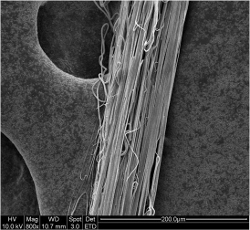
Early investigation using electrospun fibers in medical device applications is mainly as implantable scaffold. The small diameters of electrospun fibers from a few hundreds of nanometers to a few microns have been shown to facilitate cell adhesion and proliferation in numerous in vitro studies. In vivo studies have also found them to exhibit good biocompatibility. Later development in the electrospinning process has seen the construction of fibrous tubes and yarns. This expanded the potential application to sutures. There are several methods of constructing sutures from electrospinning. For finite length yarn, a rotating disk may be used to collect a bundle of fibers along the edge of the disk. Several other methods such as using water-bath as collector have been devised to collect continuous electrospun yarn. The ability to construct continuous electrospun yarn and relatively benign inflammatory response [Kashiwabuchi et al 2017] makes it an highly attractive candidate as suture.
Most electrospun sutures often includes other functional properties such as drug elution or anti-bacterial. The relative ease of loading the solution with other substances and electrospinning to form fibers make this an attractive option. Further, large surface area of small diameter electrospun fibers give greater exposure of the active substances to the surrounding. Liu et al (2010) electrospun poly(ε-caprolactone) (PCL) yarn containing ampicillin sodium salt, which is a common anti-microbial drug. The drug-loaded yarn has an initial burst release in the first hour and the release is almost completed in 96 hours. Kashiwabuchi et al (2016) constructed an electrospun poly(L-lactide) (PLLA)/ polyethylene glycol (PEG) microfibrous yarn loaded with levofloxacin targeted for ophthalmic surgery. In vitro release study showed an initial burst release for the first 48 hours followed by linear release for more than 2 months with approximately 65% cumulative release. The amount of released levofloxacin within the first 24 hrs and after 7 days were found to inhibit S. epidermidis in separate tests. In vivo tests on corneal stroma of male Sprague-Dawley rats showed no inflammation with histological results similar to that of implanted commercial nylon sutures. Weldon et al (2012) electrospun poly(lactic-co-glycolic acid) (PLGA) sutures containing anesthesia (bupivacaine HCl). With localized release of anesthesia at the operation site, it reduces postoperative discomfort and the need for other forms of anesthesia. The release of the drug in electrospun PLGA started relatively slowly and linearly for the first two days. Thereafter, the release increased rapidly from day two to day seven and all drugs were released after 12 days. Comparing the mechanical strength, electrospun drug-free PLGA suture is an order of magnitude lower than commercial PLGA suture (Vicryl, 30N).
An advantage of conventional suture made of microfilaments is its greater strength and mechanical property consistency. Electrospinning has been shown to be a very versatile nanofiber fabrication technique and several methods have been developed to deposit nanofibers over a core filament. Madheswaran et al (2022) used AC electrospinning to construct a nanofibrous sheath over a suture yarn made of either polyamide 6 or polyurethane. A polymer solution containing antibacterial chlorhexidine (CHX) was injected through a needle nozzle and electrospun. The core yarn passed across the path of the electrospinning jet and the nanofibers wrapped around the core yarn. The resultant composite yarn demonstrated antibacterial properties against Escherichia coli and Staphylococcus aureus. Tests using 3T3-SA mouse fibroblast cells showed that the composite yarn with CHX to be biocompatible.
Electrospun yarns have been tested as potential future suture material in particular, drug-loaded yarns. While the drug release performance is favourable to its intended application, more studies and modifications to the yarn is needed to improve its mechanical strength. Currently, electrospun yarns are much weaker than its commercial counterpart. Further investigations are also required to determine the benefits of having finer diameter fibers in the construction of suture over microfibers which are currently in use.
Published date: 16 May 2017
Last updated: 11 October 2022
▼ Reference
-
Kashiwabuchi F, Parikh K S, Omiadze R, Zhang S, Luo L, Patel H V, Xu Q, Ensign L M, Mao H Q, Hanes J, McDonnell P J. Development of Absorbable, Antibiotic-Eluting Sutures for Ophthalmic Surgery. Translational Vision Science & Technology 2017; 6: 1.
Open Access
-
Liu H, Leonas K K, Zhao Y. Antimicrobial Properties and Release Profile of Ampicillin from Electrospun Poly(ε-caprolactone) Nanofiber Yarns. Journal of Engineered
Open Access Fibers and Fabrics 2010; 5: 10.
-
Madheswaran D, Sivan M, Valtera J, Kostakova E K, Egghe T, Asadian M, Novotny V, Nguyen N H A, Sevcu A, Morent R, Geyter N, Lukas D. Composite yarns with antibacterial nanofibrous sheaths produced by collectorless alternating-current electrospinning for suture applications. J Appl Polym Sci 2022; 139: 51851.
-
Weldon C B, Tsui J H, Shankarappa S A, Nguyen V T, Ma M, Anderson D G, Kohane D S. Electrospun drug-eluting sutures for local anesthesia. J Control Release 2012; 161: 903.
▲ Close list
 ElectrospinTech
