Both polymeric and inorganic nanofibers membrane have been developed for the purpose of water filtration. Although the physical attributes of the membrane from both materials may be very similar, their filtration performance differs for smaller particles.
In water treatment, microfiltration is often carried out as a pre-treatment to remove particles that passes through the coagulation and sedimentation process before going into ultrafiltration, nano-filtration or reverse osmosis. Removal of microparticles enhances the lifespan and efficiency of the downstream filtration process and lower the overall treatment cost as the later filters are generally more expensive than the microfilters.
Electrospun membrane has been shown to be very effective in filtering out microparticles [Gopal et al 2006, Gopal et al 2007]. A comprehensive study on the microparticles rejection by Gopal et al (2007) using heat treated polysulfone (PSU) electrospun nonwoven membrane with bubble point of 4.6 µm showed that full flux recovery was possible for particle size 7, 8 and 10 µm with more than 99% separation factor. Full flux recovery was also reported for 5 µm size particles with separation factor greater than 90% using heat treated polyvinylidene fluoride (PVDF) membrane [Gopal et al 2006]. However, flux recovery was incomplete for particle sizes less than 3 µm. Examination of the cross section of the membrane showed that for larger particles (more than 5 µm), the membrane act as a size exclusion filter or screen filter with the particles trapped on the surface of the membrane [Gopal et al 2007]. Low adhesion force between the particles and the fiber surface meant that the flux can be recovered by stirring in the membrane module [Gopal et al 2006, Gopal et al 2007].
When the particle sizes are less than 3 µm, in particular 1 and 2 µm particles, a dense cake-layer formed on the surface of the membrane. This cake formation cause irreversible fouling of the membrane as the particles are trapped between the pores [Gopal et al 2006]. Below 1 µm, particles were able to migrate into the depth of the membrane and were trapped on the surface of the nanofibers through surface adhesion. However, separation factor for particles less than 1 µm is less than 50% [Gopal et al 2007]. Liu (2009) used cross-linked polyvinyl alcohol electrospun fibers on a polyethylene terephthalate (PET) nonwoven supporting substrate as the separation membrane for 0.2 µm particles. A similar cake formation was found on the electrospun membrane and the same was found on Millipore GSWP 0.22 µm membrane. Contrary to the results from Gopal et al (2007), initial rejection of more than 95% was achieved for the electrospun membrane. However, the permeate flux deteriorate quickly over the next 20 minutes due to the cake formation. Compared with Millipore GSWP 0.22 µm membrane, the rejection rate and permeate flux are both higher for electrospun membrane over the duration of the study (20 minutes).
Using electrospun nylon 6 membrane, Aussawasathien et al (2008) achieved similar results with close to 100% separation for 6 µm particles for membrane thickness of 0.15 mm and 0.6 mm. Cake formation was also reported for separation of 1 µm particles although the separation for 0.5 µm particles were greater at 84% for 0.15 mm thick membrane. Both hydrophobic membrane and hydrophilic membrane demonstrated similar particle separation efficiency for particles greater than 5 µm but the results for smaller particles are not in agreement. More tests need to be carried out to determine whether hydrophobicity play a part in the separation of smaller particles.
Suaste-Gómez et al (2022) investigated the use of electrospun membrane as biological sieve for the separation of yeast and bacteria. The yeasts used in the test was Candida albicans type with dimensions of 7 µm and the bacteria was Brevibacterium ravens purgense with dimensions of 1.4 µm. Electrospun polylactic acid (PLA) of thickness 66 µm, 83 µm and 128 µm were used in the study. With the thinner membranes, some yeasts were found to pass through the membrane while the 128 µm thick membrane was able to reject all the yeasts. Counterintuitively, it was reported that there was no bacteria passing through the thinnest membrane (66 µm) while the thickest membrane (128 µm) reported the most bacteria passing through. Other independent studies will be required to validate this observation. It has been reported in other studies that particles 1 to 2 µm [Gopal et al 2006; Aussawasathien et al 2008] lead to cake formation on the surface of the membrane. However, the condition used in the filtration process is different. For Suaste-Gómez et al (2022), the volume of solution containing the yeasts or bacteria that was loaded on the electrospun membrane was very small compared to the other studies and the pressure applied in this study is also much smaller than other studies on the filter performance.
To allow better handling of the microfiltration membrane, Homaeigohar (2011) uses poly(ethylene terephthalate) (PET) sublayer to support an electrospun polyether sulfone (PES) layer. Consistent with other results, the particle challenge test showed excellent rejection of microparticles with high permeability but significant cake formation for particles less than 1 µm. The study also demonstrated the importance of heat treatment of the nanofibrous membrane to maintain its integrity. None heat treated membrane was shown to suffer from compaction resulting in loss of permeability.
As a stand-alone membrane, electrospun membrane are not suitable for filtering particles less than 3 µm due to dense cake formation. Filtration performance for particles less than 1 µm is expected to be poor due to the large pore size of the membrane. However, as part of a composite membrane, electrospun fiber layer was shown to facilitate capturing of particles less than 1 µm while maintaining a high flux. Zhao et al (2013) constructed a composite membrane with a phase inversion polyvinylidene fluoride (PVDF) top layer, an electrospun polyvinylidene fluoride (PVDF) membrane intermediate layer on a nonwoven PET substrate. Polystyrene particles tested have a size of 160 nm, 500 nm and 900 nm. Stand-alone PVDF exhibits the highest flux but very low rejection with retention ratio lower than 10%. Commercial ultrafiltration shows >99.9% rejection for the larger particles but 89% for the smallest particle and a flux rate of 870 L/m2h, an order of magnitude slower than electrospun PVDF membrane. The composite membrane demonstrated rejection of more than 99.9% for all particles sizes and a much higher flux of 4900 L/m2h for the largest particle. Recovery of the composite membrane for 190 nm particles is also much better than commercial membrane. Better flux recovery of the composite membrane is attributed to its screen filtration property where particles are trapped on the surface while commercial membrane exhibits depth filtration where particles are trapped inside the membrane [Zhao et al 2013].
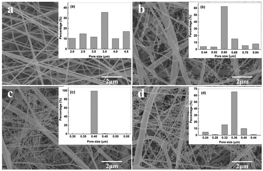
Electrospinning under suitable conditions, is able to produce nanofibrous nonwoven membrane with nanowebs between fibers. This typically involves adding salt to improve solution conductivity [Li et al 2016] or increase the electrospinning voltage [Ding et al 2006] such that secondary branches split off from the main electrospinning jet. Li et al (2016) showed that the presence of nanowebs enables the membrane to be used for microfiltration. Using electrospun polyvinylidene fluoride (PVDF) with tetrabutylammonium chloride (TBAC) added, the resultant membrane was able to reject 99.9% of 300 nm polystyrene particles and a high pure water flux of 2.88 x 10⁴L.m-2h-1 under the pressure of 25 psi. With electrospun PVDF membrane without the nanowebs, the rejection was only 46%. Despite the good rejection rate, more studies are needed to determine the drop in flux as the particles accumulates and the recovery capability of this membrane.
| Membrane description
|
Characteristics
|
Reference
|
|
Heat treated polysulfone electrospun nonwoven membrane, fiber dia. approx. 470 nm
|
Pore size: 4.6
Liquid penetration pressure: 1.8 psi
|
Gopal et al 2007
|
|
Heat treated polyvinylidene fluoride electrospun nonwoven membrane, fiber dia. approx. 380 nm
|
Pore size: 10.6 - 4 µm
Transmembrane pressure: 0.6 bar
|
Gopal et al 2006
|
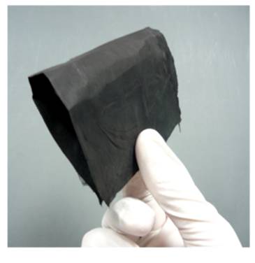
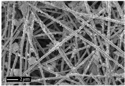
Water filtration membrane made of carbon nanofiber and carbon-silicon nanofiber (CNF/Si) was found to be very good at rejecting particles less than 100 nm compared to polymeric nanofiber filtration membranes. Faccini et al (2015) constructed CNF/Si from pyrolysis of electrospun nanofiber made of polyacrylonitrile (PAN) with tetraethoxyorthosilicate. The composite CNF/Si membrane was found to be much more flexible than pure CNF membrane. The membrane also showed very large water flux at 47620 Lm-2h-1bar-1. 95% retention was observed for Au nanoparticles of 100, 50 and 25 nm. For 10 nm particles, the filtration efficiency dropped to about 66%. However, this is much higher than the filtration efficiency for reported electrospun polymeric membrane [Gopal et al 2000]. Filtration of TiO2 nanoparticles (1- to 15 nm diameter) was also very high at 94.1% by the CNF/Si membrane. Good performance of CNF/Si membrane filter was attributed to strong electrostatic interaction of metal and metal oxide nanoparticles with the CNF/Si membranes.
Published date: 30 April 2014
Last updated: 03 January 2023
▼ Reference
-
Aussawasathien D, Teerawattananon C, Vongachariya A. Separation of micron to sub-micron particles from water: Electrospun nylon-6 nanofibrous membranes as pre-filters. Journal of Membrane Science 2008; 315: 11.
-
Ding B, Li C, Miyauchi Y, Kuwaki O, Shiratori S. Formation of novel 2D polymer nanowebs via electrospinning. Nanotechnology 2006; 17: 3685.
-
Faccini M, Borja G, Boerrigter M, Martin D M, Crespiera S M, Vazquez-Campos S, Aubouy L, Amantia D. Electrospun Carbon Nanofiber Membranes for Filtration of Nanoparticles from Water. Journal of Nanomaterials 2015; 2015: 247471.
Open Access
-
Gopal R, Kaur S, Ma Z, Chan C, Ramakrishna S, Matsuura T. Electrospun nanofibrous filtration membrane. Journal of Membrane Science 2006; 281: 581.
-
Homaeigohar S S. Functional Electrospun Nanofibrous Membranes for water filtration. PhD Thesis 2011, University of Kiel.
Open Access
-
Li Z, Kang W, Zhao H, Hu M, Wei N, Qiu J, Cheng B. A Novel Polyvinylidene Fluoride Tree-Like Nanofiber Membrane for Microfiltration. Nanomaterials 2016; 6: 152.
Open Access
-
Liu Y. High-Flux Microfiltration Filters Based on Electrospun PVA Nanofibrous Mats. Master of Science Thesis 2009, Stony Brook University.
Open Access
-
Suaste-Gómez E, Rodríguez-Roldán G, Pérez-Solis I, Torres-Huerta A, Cruz-Cruz C, Tapia-Ramírez J. Electrospinning Polylactic Acid Polymer Membranes as Biological Sieve for Yeast and Bacteria. Materials Sciences and Applications 2022; 13: 389.
Open Access
-
Zhao Z, Zheng J, Peng B, Li Z, Zhang H, Han C C. A novel composite microfiltration membrane: Structure and performance. Journal of Membrane Science 2013; 439: 12.
▲ Close list