Home | About | Contact


Mineralisation of Electrospun fibers


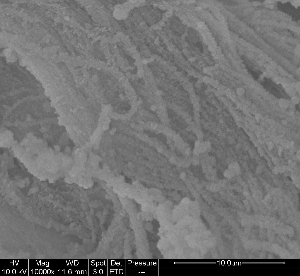
SEM image showing mineral particles deposit on the nanofibers using alternate dipping method.

Electrospun fibers have been widely investigated for use as tissue regenerative scaffold. For scaffold targeted for bone regeneration, having calcium ions especially calcium phosphate minerals are known to facilitate bone regeneration and integration. This has led to researchers developing or utilizing different methods to introduce calcium ions or its minerals to electrospun scaffolds.

Perhaps the most direct method is to mix calcium salt or nanoparticles into the solution and electrospinning. With this, it is possible to add calcium to any solution that can be electrospun to give fibers. One of the earliest studies of incorporating calcium ions into electrospun fibers is by Fujihara et al (2005). Using a blend of calcium carbonate and polycaprolactone (PCL), Fujihara et al (2005) showed that the resultant electrospun membrane was able to support osteoblast attachment and proliferation. Later, hydroxyapatite, the dominant form of calcium found in bones, have been blended into polymer solution for electrospinning into fibers. To ensure uniform distribution of calcium phosphate nanoparticles in electrospun fibers, Panzavolta et al (2016) used a triaxial nozzle to spontaneously fabricate calcium phosphate nanoparticles and drawing them along the core of electrospinning gelatin. In the triaxial nozzle, Calcium chloride dihydrate (CaCl22H2O) and sodium phosphate monobasic dodecahydrate (Na3PO412H2O) solution was loaded into the inner core and intermediate cylinder respectively while the outermost cylinder was loaded with gelatin solution. During electrospinning, all three solutions were ejected concurrently. As CaCl22H2O and Na3PO412H2O reacted to form calcium phosphate solution at the inner portion of nozzle, gelatin solution at the outermost cylinder was drawn from the nozzle tip by electrospinning and bringing with it the formed calcium phosphate nanoparticles in its core.

Nano-hydroxyapatite may come in the form of spherical particles, whiskers and rods. Haider et al [2014] compared obsteoblast response to poly(lactide-co-glycolide) nanofiber embedded with spherical HA nanoparticles and nanorod HA and found that the later HA morphology showed significantly higher cell adhesion, proliferation and osteogenesis performance. It was hypothesized that the difference in obsteoblast response is due to difference in the charges on rod and spherical shape HA particles. A rod shape HA will carry more positively charged particles which favours adsoprtion of negatively charged acidic protein while spherical HA carries more positive charges. Nevertheless, both rod and spherical HA embedded nanofibers perform better than neat poly(lactide-co-glycolide) nanofibers. A limitation to this method is the amount of calcium that can be loaded due to its solubility in the solution or aggregation for nanoparticles. The calcium minerals would also be embedded in the polymer matrix which reduces its exposure to cells.

There are several methods to load electrospun fibers with calcium minerals on its surface. Tavakoli-Darestani et al (2013) simply coated electrospun poly (lactide-co-glycolide) (PLGA) with collagen followed by soaking the coated fibers in a suspension of nano-HA particles overnight. The adhesion of the nano-HA particles on the surface of the collagen coated PLGA fibers were found to be strong enough to withstand washing. Soaking a material in simulated body fluid (SBF) is a common technique to deposit hydroxyapatite (HA) . However, most man-made polymer material such as PCL and polylactic acid (PLA) does not have favourable reactive site for HA deposition. Electrospun PCL polymer fibers containing HA nanoparticles have been shown to encourage HA deposition on fiber surface in SBF while there are no HA deposition without HA nanoparticles preloaded [Hassan et al 2014]. Similarly, where amorphous calcium phosphate (ACP) nanoparticles were loaded into electrospun polylactic acid fibers, mineralization on the surface of the fibers were already observable in a day when soaked in SBF. Without ACP, no mineral deposits were seen on pure PLA fibers after 7 days in SBF [Fu et al 2016]. Soaking a scaffold in SBF is known to deposit stable HA nanoparticles but this process typically takes a few days. A faster method is to produce HA on a scaffold surface is to use chemical reaction between calcium and phosphate salts.

The right combination and composition of calcium and phosphate salt typically produces HA nanoparticles in minutes. A simple way is to dip the scaffold in calcium solution followed by phosphate solution. The soaking process is alternated between the two solutions until the desired amount of HA is produced. Ngiam et al (2009) demonstrated the feasibility of using this method to deposit HA nanoparticles on electrospun fibers of poly(L-lactic acid) (PLLA) and poly(L-lactic acid)/collagen (PLLA/Col). While HA nanoparticles were deposited on both materials, it was the PLLA/Col nanofibers that showed faster and more uniform HA particles distribution. This is probably due to the presence of functional groups on collagen that facilitates calcium and phosphate deposition. The alternate dipping method has been used to deposit HA nanoparticles in electrospun nanofibrous blocks [Teo et al 2011].


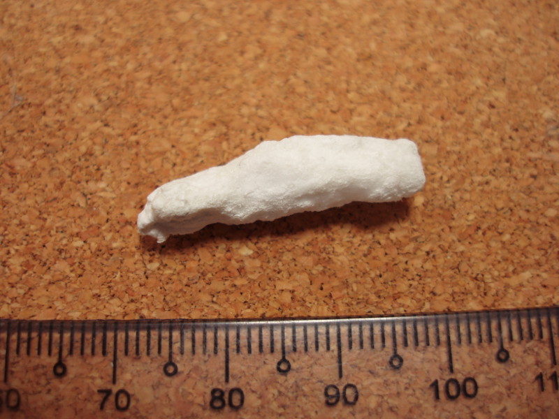
Mineralized electrospun scaffold using alternate-dipping method.

Published date: 17 January 2017
Last updated: 24 October 2017

Comments
[+]


Google Ad.