Electrospun scaffold pore size has a significant influence on the ability of the cells to infiltrate into the scaffold. In most applications, it is preferred that cells are able to infiltrate into the scaffold for better integration with the surrounding host tissue. Studies have also suggested that larger pore size resulted in better cell proliferation although this may be due to greater space available for cell proliferation in larger pore size scaffold. For scaffold with smaller pore size, only the surface of the scaffold is available for cells to proliferate while a larger pore size scaffold allows cells to occupy the depth of the scaffold. In a study by Soliman et al (2011), electrospun polycaprolactone (PCL) scaffold with different fiber diameter and packing density was carried to determine its effect on human umbilical vein endothelial cells (HUVECs) proliferation. Scaffold with higher packing density was induced by using an auxiliary ring electrode to focus the electrospinning jet to a smaller deposition area. In general, for both microfibers and nanofibers, scaffold with lower packing density showed better cell proliferation and infiltration. Scaffold comprising of microfibers and lower packing density give the best cell viability and proliferation rate. However, smaller diameter fibrous membrane has been shown to be better for cell differentiation and proliferation for other cell type [Sisson et al 2010].
Physical displacement of nanofiber during electrospinning through the use of air-flow has also been shown to increase the pore size of electrospun membrane. This method works by having a perforated mandrel collector and passing air through the perforations as the fibers are collected on it. Using this technique with different air pressure, McClure et al (2012) was able to fabricate electrospun polycaprolactone (PCL) tubes with maximum average pore size of 7.6 µm. Electrospun PCL tube fabricated on solid tube has an average pore size of 3.8 µm. In their experiment, the pore size increases with increasing air pressure but starts to reduce once it exceeds the optimum air pressure of 100 kPa. Cell infiltration using fibroblast showed infiltration depth of 186 µm for the largest average pore size scaffold compared to 21 µm for scaffold fabricated on solid mandrel. The use of air flow from the collector was able to increase the pore size of the membrane after optimization of the flow conditions. However, more studies are necessary to determine factors that affect fiber displacement in the creation of the pore and the influence of the air flow as the membrane thickness increases. Cell cultures on the scaffold although showed significant improvement in cell infiltration, does not demonstrate cell migration throughout its depth [McClure et al 2012].
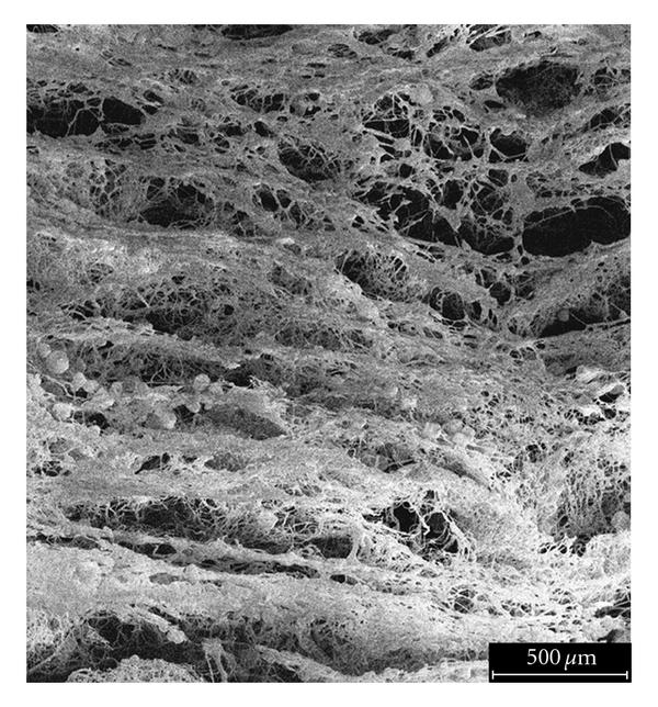
Temporary spacers are the most commonly used method of increasing the pore size of the membrane. Spaces used include particles, sacrificial fibers or ice crystals. Wulkersdorfer et al (2010) alternate electrospinning of poly(glycolic acid) and spraying of sucrose-ethanol suspension with sucrose particle size from 250 to 350 µm, at time interval of 1 min, 2 min and 10 min. At interval of 2 min, pore size of 150 µm was formed with macroporous interconnections. However with 10 min interval, layers of dense electrospun fibers with distinct space between the layers were formed despite having larger average pore size of more than 300 µm. Cell infiltration study showed good penetration in the electrospun membrane with pore size generated by 2 min interval but the membrane from 10 min interval showed poor penetration.
Given that particles only takes up isolated space within the membrane, the interconnection between resultant pores may be reduced if the particles distribution are sparse. A better way to ensure interconnected pores is to use sacrificial fibers that are evenly dispersed between the desired electrospun fibers. Frey et al (2007) used polyamide-6 as the primary electrospun fiber and simultaneously electrospin poly(ethylene oxide) as the sacrificial fibers. With just polyamide-6 fibers or without removing the sacrificial fibers, the pore size distribution is relatively narrow with the majority pore size below 200 nm diameter. After removal of the sacrificial fibers, the pore size distribution significantly increases with pore diameter ranging from 100 nm to a micron and 50% of the pores larger than 200 nm in diameter. Similarly, Guimaraes et al (2010) simultaneously electrospin polycaprolactone fiber and polyethylene oxide fiber onto a collector with the latter used as a sacrificial material. After removal of the sacrificial fiber, the pore size of the membrane increases from about 2.5 µm to 5 µm. Culturing osteoblast on both scaffolds showed cell penetration through the electrospin polycaprolactone fiber membrane with the sacrificial membrane removed while cells remained only on the surface of polycaprolactone membrane.
Having pores is not the only way to create greater surface area on a membrane. An alternative is to have the undulating surfaces. Patterned membranes comprising of nanofibers featuring micro and macro troughs can be easily created by depositing the electrospun fibers on a patterned collector. Cell culture comparing proliferation of mouse osteoblastic cell between patterned/textured nanofiber and randomly oriented nanofiber showed faster proliferation on the textured nanofiber membrane. However, initial cell adhesion is better on randomly oriented nanofiber membrane [Wang et al 2009]. Neves et al (2007) has observed that osteoblastic cells showed preferential adhesion to the regions of randomly oriented nanofibers and this may explain the lower cell numbers recorded by Wang et al (2009) on day 1 for patterned nanofiber mesh compared to unpatterned, randomly oriented nanofiber membrane. Patterned nanofibrous membrane was also shown to have varying pore sizes between the fibers deposited on the wire and the space between. Larger pore size in the space between has been shown to faciliate fibroblast migration into the depth of the membrane compared to the thicker segments where the cells tend to remain on its surface [Vaquette et al 2011].
Another way of influencing fiber packing density is by having the fibers embedded in a hydrogel matrix. This can be easily constructed by immersing the fibers in hydrogel precursor prior to gelation. Chopping electrospun fibers into shorter strands will allow better distribution of the fibers throughout the hydrogel if they can be separated into individual strands. Rivet et al (2015) fabricate individual short strand nanofibers by collecting aligned fibers on a substrate with a thin film of polyvinyl alcohol (PVA) as release agent. The substrate with the deposited aligned fibers was cut into short segments of about 1 mm width, perpendicular to the fiber orientation. The PVA was subsequently removed from the fibers by dissolving in water. The dispersed chopped fibers were finally mixed in hydrogel.
Published date: 18 October 2016
Last updated: 02 March 2021
▼ Reference
-
Frey M W, Li L. Electrospinning and Porosity Measurements of Nylon-6/Poly(ethylene oxide) Blended Nonwovens. Journal of Engineered Fibers and Fabrics 2007; 7: 31.
Open Access
-
McClure M J, Wolfe P S, Simpson D G, Sell S A, Bowlin G L. The use of air-flow impedance to control fiber deposition patterns during electrospinning. Biomaterials 2012; 33: 771.
-
Neves N M, Campos R, Pedro A, Cunha J, Macedo F, Reis R L. Patterning of polymer nanofiber meshes by electrospinning for biomedical applications. Int. J. Nanomed. 2007; 2: 433.
-
Rivet C J, Zhou K, Gilbert R J, Finkelstein D I, Forsythe J S. Cell infiltration into a 3D electrospun fiber and hydrogel hybrid scaffold implanted in the brain. Biomatter 2015; 5: e1005527.
Open Access
-
Sisson K, Zhang C, Farach-Carson M C, Chase D B, Rabolt J F. Fiber diameters control osteoblastic cell migration and differentiation in electrospun gelatin. J Biomed Mater Res Part A 2010; 94A: 1312.
-
Soliman S, Sant S, Nichol J W, Khabiry M, Traversa E, Khademhosseini A. Controlling the porosity of fibrous scaffolds by modulating the fiber diameter and packing density. Journal of Biomedical Materials Research A 2011; 96A: 586.
-
Vaquette C, Cooper-White J J. Increasing electrospun scaffold pore size with tailored collectors for improved cell penetration. Acta Biomaterialia 2011; 7: 2544.
-
Wang Y, Wang G, Chen L, Li H, Yin T, Wang B, Lee J C M, Yu Q. Electrospun nanofiber meshes with tailored architectures and patterns as potential tissue-engineering scaffolds. Biofabrication 2009; 1: 015001.
-
Wulkersdorfer B, Kao K K, Agopian V G, Ahn A, Dunn J C, Wu B M, Stelzner M. Bimodal Porous Scaffolds by Sequential Electrospinning of Poly(glycolic acid) with Sucrose Particles. International Journal of Polymer Science 2010; 436178: 9 pages.
Open Access
▲ Close list