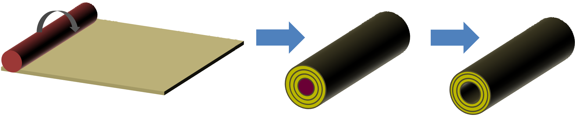
An indirect method of constructing a nanofibrous tube is by rolling a nanofibrous membrane over a rod. A polymer coating may be applied over the surface to prevent the unraveling of the membrane. This polymer coating may also function as a barrier to control drug or biomolecule release from the nanofibrous membrane [Madduri et al 2010]. While this method requires two steps to construct a tube excluding the application of a bonding agent, it does offer some advantages. The removal of the constructed tube from the inner rod template is easier compared to tubes formed from direct deposition of fibers on a rod. A thinner wall tube may also be constructed using this method. A limitation of electrospun membrane as a tissue scaffold is that the small pore sizes between adjacent fibers limits cell migration into its depth. Seeding and culturing cells on the electrospun membrane prior to rolling into the form of a tube helps to introduce cells across the thickness of the tube wall. This method has been demonstrated by Wang et al (2016) to construct blood vessel-mimicking tissues. To prevent unravelling of the tube, fibrin glue was used to keep the rolled up electrospun membrane in place. This allows the construction of a blood vessel tubular scaffold with cells within 70 minutes. For membrane that is made of gelatinous material, additional adhesive agent may not be necessary if the rolled tube is to be used in a wet environment. Zhou et al (2016) electrospun a polycaprolactone/gelatin (PCL/gel) membrane for seeding of smooth muscle cells (SMC). After 7 days of culture, the membrane with the cells was rolled over a silicone tube for further culture in a pulsatile bioreactor. The membrane was degraded after 6 to 8 weeks of culture but still maintained sufficient integrity in the form of a tube for the underlying supporting silicone tube to be removed.
Constructing a tube with different layers and fiber alignment will depend on the fiber alignment of the membrane. This may be easier to control than getting the fibers to align when depositing them directly on the rod.
The variety of tubes in terms of its surface structure and fiber distribution and orientation that can be constructed using the roll on method is also much greater. Kim et al (2016) constructed an electrospun tube with areas of high transparency and low transparency. Higher transparency regions are made out of aligned fibers while randomly oriented fibers form the low transparency regions. Sectors comprising of highly aligned nanofibers were created by having non-conductive cellophane tape on the rotating drum surface. The non-conductive cellophane tape creates a "gap" within a more conductive surface and this aligned fibers formed across the width of the cellophane tape similar in principle to the parallel electrodes collector for collecting aligned fibers. The sectors comprising of oriented fibers have much greater transparency than the other sectors which is made out of randomly oriented fibers. By deliberately specifying areas of higher transparency, they were able to roll the mat into a tube with sections that has greater transparency. This may facilitate nerve reconstructive surgery by having both edges of the tube more transparent.
The roll-on rod may also be used to introduce luminal structure into the conduit. Zennifer et al (2025) used a combination of 3D-printed thermoplastic polyurethane (TPU) fiber lattice and electrospun poly(3-hydroxybutyrate-co-3-hydroxyvalerate) (PHBV) (PHBV) fibers to first form a flat sheet. The sheet is subsequently rolled tightly using a steel rod and heat sealed to form a conduit. An advantage of using 3D printing is that the infill density of the grid structure can be controlled. The electrospun fibers were deposited directly on the 3D printed grid. Aligned fibers were obtained by electrospinning on the printed grid mounted on a rotating drum. To enhance contact guidance of the peripheral nerve, the electrospun aligned fibers were oriented along the length of the conduit. In vivo tests using 10 mm sciatic nerve defect in Wistar rats treated with the conduit showed muscle innervation and axon healing comparable to autografts over 4 months.
▼ Reference
-
Kim J I, Hwang T I, Aguilar L E, Park C H, Kim C S. A Controlled Design of Aligned and Random Nanofibers for 3D Bi-functionalized Nerve Conduits Fabricated via a Novel Electrospinning Set-up. Scientific Reports 2016; 6: 23761.
Open Access
-
Madduri S, Papaloizos M, Gander B. Trophically and topographically functionalized silk fibroin nerve conduits for guided peripheral nerve regeneration. Biomaterials 2010; 31: 2323.
-
Wang N, Tang L, Zheng W, Peng Y, Cheng S, Lei Y, Zhang L, Hu B, Liu S, Zhang W, Jiang X. A strategy for rapid and facile fabrication of controlled, layered blood vessel-like structures. RSC Adv. 2016; 6: 55054.
-
Zennifer A, Kumar S K P, Bagewadi S, Unnamalai S, Chellappan D, Abdulmalik S, Yu X, Sethuraman S, Sundaramurthi D, Kumbar S G. Innovative spiral nerve conduits: Addressing nutrient transport and cellular activity for critical-sized nerve defects. Bioactive Materials 2025; 44; 544.
https://www.sciencedirect.com/science/article/pii/S2452199X2400481X Open Access.
-
Zhou R, Zhu L, Fu S, Qian Y, Wang D, Wang C. Small Diameter Blood Vessels Bioengineered From Human Adipose-derived Stem Cells. Scientific Reports 2016; 6: 35422
Open Access
▲ Close list