In water treatment, electrospun membrane is very effective as microfilters and they are able to remove bacteria by size exclusion and inactivation. However, a non-polluted water source may still harbour viruses which are able pass through electrospun membrane due to their size which may be as small as 20 nm. In the absence of high pressure system, reduction of electrospun membrane pore size to the nanometer scale to separate virus is impractical as especially if it is designed for rural or outdoors usage. The variety of material that can be electrospun means that filtration by electrospun membrane need not be restricted to size exclusion. Functionalization of the fiber may be used to incorporate surface adsorption capability for removal of unwanted substances from the water. Anti-viral substances may also be added to the electrospun fibers.
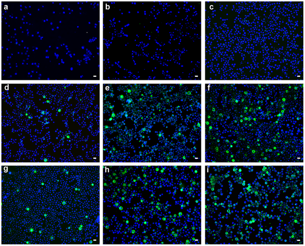
Most viruses such as MS2 bacteriophage have a negatively charged surface in a neutral solution [Michen and Graule 2010] and this enables their removal using adsorption. Functionalizing of electrospun fibers to give a positive surface charge will allow its membrane to remove virus using this method while retaining its high flux and low pressure drop property. Wang et al (2013) demonstrated the importance of using electrostatic interactions for virus removal using polyacrylonitrile fiber infused with carboxylated cellulose nanofibers with and without grafted polyvinylamine (PVAm). Fiber with grafted PVAm showed excellent adsorption efficiency for MS2 while the adsorption dropped dramatically without grafting as carboxylate is negatively charged. Bai et al (2013) used porcine parvovirus (PPV) which has diameter of less than 25 nm as the model to demonstrate virus removal using electrospun membrane. Electrospun chitosan nanofiber which has a positive charge was first tested for removal of negatively charged porcine parvovirus (PPV). However, only 30% of the PPV was removed. To improve the binding efficiency of chitosan nanofibers, quaternary amine bonded to it and a binding efficiency of 70% was achieved. Addition of graphene and polyvinyl alcohol to quaternized chitosan further improves the binding efficiency of PPV to more than 90%. While it has been suggested that hydrophobicity of graphene has contributed to the improved binding, it is unclear why the addition of PVA also improves binding since PVA is not hydrophobic. Filtration characteristic for virus is consistent to filtration of other particles. Mi (2013) showed that increasing the layers of nanofibrous membranes, the filtration efficiency increases.
Anti-viral substances may also be incorporated into electrospun fibers using simple technique like blending. Lhotakova et al (2012) prepared polycaprolactone and polyurethane (Tecophilic®)nanofibers with an encapsulated 5,10,5,20-tetraphenylporphyrin photosensitizer for virus inactivation. The photosensitizer was able to generate O2(1Δg) which causes oxidative damages to biological materials. When tested against non-enveloped mouse polyomavirus and enveloped baculovirus in infected cells, the number of infected cells reduces significantly upon exposure to irradiation over the experimental duration of 30 minutes.
Published date: 14 July 2015
Last updated: -
▼ Reference
-
Bai B Y, Mi X, Xiang X, Heiden P A, Heldt C L. Non-enveloped virus reduction with quaternized chitosan nanofibers containing graphene. Carbohydrate Research 2013; 380: 137.
-
Lhotakova Y, Plistil L, Moravkova A, Kubat P, Lang K, Forstova J, Mosinger J. Virucidal Nanofiber Textiles Based on Photosensitized Production of Singlet Oxygen. PLoS ONE 2012; 7: e49226.
Open Access
-
Mi X. Electrospun Quaternized Chitosan Fibers for Virus Removal from Drinking Water. Master of Science Thesis, Michigan Technological University 2013.
Open Access
-
Michen B, Graule T. Isoelectric points of viruses. Journal of Applied Microbiology 2010; 109: 388.
-
Wang R, Guan S H, Sato A, Wang X, Wang Z, Yang R, Hsiao B S, Chu B. Non-enveloped virus reduction with quaternized chitosan nanofibers containing graphene. Journal of Membrane Science 2013; 446: 376.
▲ Close list