
▼ Reference
- Biazar E, Keshel S H, Pouya M. Efficacy of nanofibrous conduits in repair of long-segment sciatic nerve defects. Neural Regeneration Research 2013; 8: 2501. Open Access
- Dinis TM, Vidal G, Jose RR, Vigneron P, Bresson D, et al. (2014) Complementary Effects of Two Growth Factors in Multifunctionalized Silk Nanofibers for Nerve Reconstruction. PLoS ONE 9(10): e109770. doi:10.1371/journal.pone.0109770 Open Access
- Ghasemi-Mobarakeh L, Prabhakaran M P, Morshed M, Nasr-Esfahani M H, Ramakrishna S. Electrical Stimulation of Nerve Cells Using Conductive Nanofibrous Scaffolds for Nerve Tissue Engineering. Tissue Engineering Part A 2009; 15: 3605
- Koh H S. Polymeric Nanofiber Conduits for Peripheral Nerve Regeneration. PhD Thesis, National University of Singapore 2009.
- Leach M K. Biomimetic Electrospun Fibers for Peripheral Nervous System Repair. University of Michigan, PhD Thesis 2013.
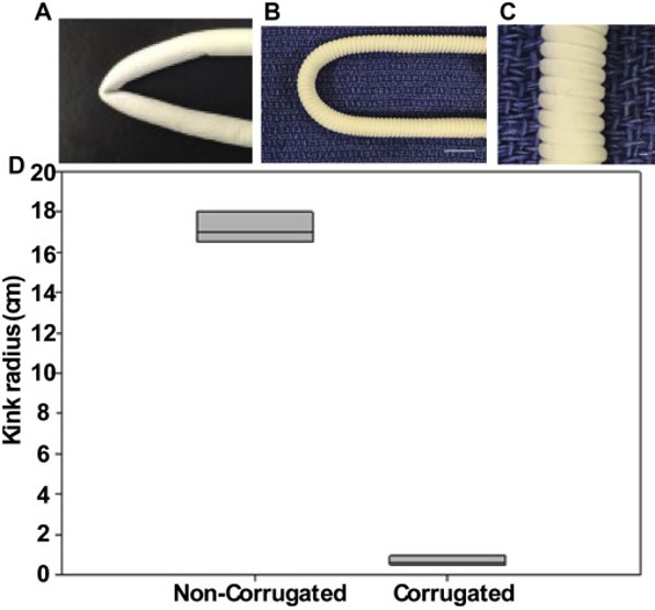
- Matsushita H, Inoue T, Abdollahi S, Yeung E, Ong C S, Lui C, Pitaktong I, Nelson K, Johnson J, Hibino N. Corrugated nanofiber tissue-engineered vascular graft to prevent kinking for arteriovenous shunts in an ovine model. JVS: Vascular Science 2020; 1: 100. Open Access
- Schaub N J, Le Beux C, Miao J, Linhardt R J, Alauzun J G, Laurencin D, Gilbert R J. The Effect of Surface Modification of Aligned Poly-L-Lactic Acid Electrospun Fibers on Fiber Degradation and Neurite Extension. PLoS ONE 2015; 10(9): e0136780. Open Access Open Access
▼ Credit and Acknowledgement
Author
Wee-Eong TEO View profile
Email: weeeong@yahoo.com
