Electrospun mesh with its high-surface area, porosity and flexibility in material selection and additives loading makes it attractive for use as wound dressing [Abrigo et al 2014]. Factors such as fluid update ability and water vapor transmission rate are some parameters that affect the performance of wound dressing through maintaining a moist environment while removing excess exudates. Wound dressings that are highly flexible are also useful for wounds on "difficult" anatomical sites. These properties are in turn influenced by material selection, structure and form of the dressing [Jones et al 2006]. The typical electrospun output comes in the form of a flat nonwoven film and its flexibility allows it to be used in most wound sites. Electrospinning may also be combined with other manufacturing processes to tailor the properties of the wound dressing. Al Kayal (2020) constructed a bilayered fibrin/poly(ether)urethane (PEU) scaffold with PEU phase-inversed membrane as the base supporting the electrospun layer of fibrin fibers. After electrospinning of fibrin, polymerization was obtained by thrombin solution spraying onto it. Adhesion of the fibrin fibers on the PEU base was found to be strong.
General Performance
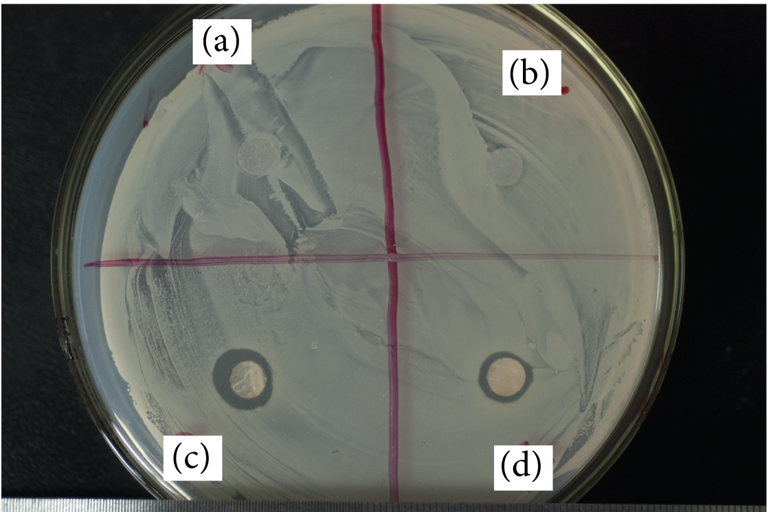
The first requirement of wound dressing is to maintain a favourable environment for healing. This involves maintaining a moist environment while removing excess exudates. Wang et al (2015) tested the performance of electrospun poly(ethylene-co-vinyl alcohol) (EVOH) on its fluid update ability and water vapor transmission rate (WVTR). Using distilled water, the fluid update of electrospun EVOH was found to be more than 80% while that of cotton gauze is about 68%. This shows that electrospun EVOH has a higher potential to cope with more exudates compared with cotton gauze. In terms of water vapor transmission rate (WVTR), it has been suggested that WVTR between 2000 g.m-2.24h-1 and 2500 g.m-2.24h-1 is preferred [Queen et al 1987]. Distilled water is commonly used in these experiments and a comparative study by Queen et al (1987) showed that the difference in WVTRs from experiment conducted using water and plasma is insignificant. With electrospun poly(ethylene-co-vinyl alcohol) (EVOH), the WVTRs were from 900 to 1000 g.m-2.24h-1 [Wang et al 2015] which is lower than the recommended WVTR. Since the selection of material will have a significant influence on WVTR, Motealleh et al (2014) used a composite of electrospun poly(e-caprolactone)/polystyrene (PCL/PS) instead. Its WVTR was found to be 1896 g.m-2.24h-1 which is very close to the recommended WVTR. On the other hand, electrospun poly(e-caprolactone) loaded with 2% tetracycline hydrochloride showed WVTR in the range of 3290-3378 g.m-2.24h-1 [Chellamani et al 2014].
Several studies have shown that cell migration is faster on oriented fibers than cast films [Shang et al 2010, Liu et al 2010]. In wound dressing, it is advantages for quicker cell coverage on the wound to facilitate recovery. Shin et al (2019) used a modified electrospinning setup to create radially patterned polycaprolactone (PCL) nanofibers to encourage faster migration of human bone marrow stem cells from the periphery to the centre of the scaffold. Comparison of cell migration speed was made with randomly oriented electrospun PCL nanofibers. Both radially and randomly oriented nanofibers have a diameter of about 380 nm and interfiber pore size of about 12 nm. From the SEM images of the scaffolds, the radial patterning of the nanofibers are not very clear. However, there are distinct difference in the migration speed of the cells on the radially patterned and randomly oriented nanofibers scaffolds. From day 5 of the cell culture, there is a difference in the cell coverage on the scaffolds. On day 7, the randomly oriented nanofiber scaffold showed a median cell-free area of 84% and the radially patterned nanofibrous scaffold showed a median cell-free area of 49%. This clearly showed the benefits of having radially patterned nanofibers in guiding the cells towards the centre of the scaffold.
Drug Release
In wound dressing for more serious cases and inflammatory skin disorders, the ability to encapsulate drugs into the dressing will enhance healing and treatment of the injured site. Brooker et al (2021) used electrospun poly(ethylene oxide) (PEO) fibres for encapsulating antioxidant and anti-inflammatory nanoparticles composed of crosslinked poly(propylene sulfide) (PPS). The electrospinning was carried out in an aqueous medium since PEO is a water soluble polymer. This electrospun PEO/PPS-NPs membrane is targeted for dry wound disorder hence the release rate of the drug will be slow and the wound dressing will have a moisturising effect. Up to 14.3% PPS-NPs has been loaded into PEO solution and electrospun into fibers with average fiber diameters of 191 nm and 166 nm for neat fibers. The PEO/PPS-NPs was found to be biocompatible when tested on human dermal fibroblasts. The PPS-NPs from the electrospun fibers were also found to retain its anti-inflammatory properties as demonstrated by TNF-α inhibition from lipopolysaccharides stimulated RAW264.7 macrophages.
Yue et al (2022) used electrospinning to encapsulate liposomes prepared with astragaloside IV (AS), and astragalus polysaccharide (APS) in polyvinyl alcohol (PVA) fibers. Both AS and APS were extracted from Astragalus membranaceus, a major herbal remedy for diabetic foot ulcers used in traditional Chinese medicine. Preparation of the solution for electrospinning is through blending of the active ingredients into the PVA solution. Efficacy of the membrane was tested in vivo on SD rats with full thickness wound. PVA membrane loaded with AS liposome (ASL) and APS was found to have less wound inflammation, greater deposition of collagen and regeneration of epithelium compared to pure PVA membrane and negative control. At day 15, the wound was completely covered in the groups with ASL/APS/PVA membrane and APS/PVA. The group with ASL/APS/PVA membrane showed the best recovery with the most closely arranged collagen fiber deposition.
Gürtler et al (2024) constructed an electrospun wound dressing containing two anti-inflammatory ingredients, salicylic acid for short term burst release and hydrocortisone for more delayed release. The wound dressing was made of three layers of electrospun polycaprolactone (PCL) membrane. The bottom layer of polycaprolactone (PCL) fibers was loaded with salicylic acid, the middle electrospun PCL layer was loaded with hydrocortisone and the top layer was pure PCL fibers without any active ingredients. The layer-by-layer wound dressing showed a 70% salicylic acid content release within the first hour while only 40% of hydrocortisone was released in the same period. 80% of hydrocortisone was released after 3 days and 90% at day 7. The delayed release has been attributed to the upper and lower layers which function as a barrier for the PBS release medium to permeate. Tests on human skin biopsies for 48 h using moistened layered PCL wound dressing demonstrated a reduction in cytokine IL-6 compared to a control sample of drug-free PCL and combination of salicylic acid ointment and hydrocortisone cream.
Bacteria Exclusion
Typical pore size of electrospun fibrous membrane is less than 5µ and this is able to prevent bacteria from reaching the wound. To demonstrate the effectiveness of electrospun layer in filtering out microbes and bacteria, Lev et al (2012) first electrospun a layer of polyurethane over a polypropylene supporting substrate. With a electrospun nanofibers weight of 3.8 g/m2, it is able to filter out E. coli although at weight less than this, E. coli was able to get pass the membrane. Chaudhary et al (2014) used an electrospun polyacrylonitrile-silver composite filter media to cover a nutrient media in room condition and passes ambient air through the filter media. When compared to the negative control which is without the protective filter media, the nutrient media protected by the nanofibrous filter remains free of bacteria growth after two months while the unprotected nutrient media show microorganism growth.
Anti-bacterial
In wound dressing application, it is essential that factors that give rise to its anti-bacterial property must not compromise wound healing. Cai et al (2010) fabricated chitosan/silk fibroin composite nanofibers using electrospinning and tested for its anti-bacterial property and biocompatibility. While the composite material showed inhibition effect on E. coli, it does not seem to be effective against S. aureus. Biocompatibility study using murine fibroblast showed that the composite membrane promotes cell adhesion and proliferation.
To prevent wound infection, the use of antibiotics is the most direct method. However, frequent use of antibiotics have led to increasing antibiotic resistance. Other methods of preventing infections have been explored to reduce the reliance of antibiotics. Bao et al (2022) used a combination of graphene (Gr) and polyphenolic tannic acid (TA) in electrospun hyaluronic acid (HA) for the construction of a wound dressing. Gr is thought to exhibit antibacterial effects by physical puncturing and cutting, oxidative stress-induced damage, and photothermal effects. TA is also known to exhibit strong antibacterial properties and crosslink with Gr through hydrogen bonding or ions. Further TA can inhibit hyaluronidase activity, to enhance the stability of hyaluronic acid To create the antibacterial wound dressing, TA and Gr were added to a solution of HA and electrospun to form the membrane. The resultant membrane undergoes photocrosslinking using a solution of 2-hydroxy-4'-(2-hydroxyethoxy)-2-methylpropiophenone and UV irradiation. When tested against Staphylococcus aureus, HA/Gr membrane has an inhibition rate of 38% while pure HA membrane has an inhibition rate of only 8%. HA/Gr/TA membrane has the highest inhibition rate of 97%. Therefore, while Gr exhibits antibacterial property, the addition of TA is necessary to bring the antibacterial effect to over 90%. Biocompatibility tests were carried out using NIH-3T3 cells on a composition of HA/Gr/TA (20% w/v HA + 0.1% w/v Gr + 0.3% w/v TA) and compared with the control which were cells cultured in the well without any membrane. Their results showed that the proliferation of the cells on the HA/Gr/TA membrane was significantly better than the control over a 36 h period which provided evidence that the Gr at such low concentration may be safe.
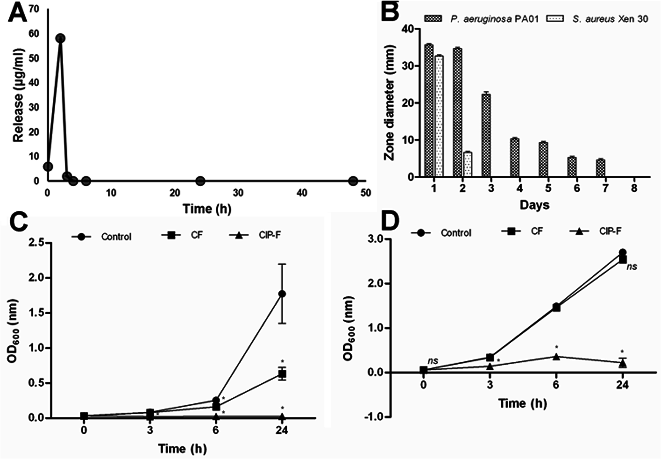
Commercially available drugs may be loaded into electrospun fibers to introduce anti-bacterial property while ensuring biocompatibility. Unnithan et al (2012) loaded polyurethane/dextran nanofiber mats with ciprofloxacin HCl (CipHCl). The resultant membrane was found to inhibit both gram-positive bacteria (S. aureus, B. subtilis) and gram-negative bacteria (E. coli, S. typhimurium, V. vulnificus. The membrane was also biocompatible with good proliferation demonstrated by culturing 3T3-L1 fibroblasts. Chellamani et al (2014) showed that wound healing from electrospun polycaprolactone (PCL) loaded with tetracycline hydrochloride was faster than common wound dressing procedure. With 2% tetracycline hydrochloride loading into the electrospun membrane, bacterial reduction was 100% on S aureus and K. pnenmonial. In a further study by Ahire et al (2015) on ciprofloxacin (CIP) eluting nanofibers, blended poly(D,L-lactide) (PDLLA) and poly(ethylene oxide) (PEO) nanofibers were used as the carrier material and tested against Pseudomonas aeruginosa and Staphylococcus aureus. Both bacteria are known to form biofilms and are commonly found in hospital acquired infections. All CIP was released from the PDLLA/PEO nanofibers within the first 3 hours. However, growth of P. aeruginosa continued to be inhibited for seven consecutive days although inhibition of S. aureus is only for 48 hours. Breast epithelial cells MCF-12A cultured on CIP loaded nanofibers were found to be viable thus demonstrating biocompatibility of the membrane.
A: Release of ciprofloxacin (CIP) from PDLLA: PEO nanofibers immediately after immersion into PBS, and 2, 3, 4, 6, 24 and 48 h thereafter, B: in vitro antimicrobial activity of CIP-containing nanofibers (CIP-F) against P. aeruginosa PA01 and S. aureus Xen 30, C: cell density of P. aeruginosa PA01 exposed to nanofibers without CIP (CF) and D: cell density of S. aureus Xen 30 exposed to CF and CIP-F. The control was without nanofibers and without CIP. Data points represent an average reading recorded from three disks per time point and three independent experiments (mean ± standard deviation). * p < 0.05. ns: not significant.
[Ahire et al PLoS ONE 2015; 10: e0123648. This work is licensed under a Creative Commons Attribution 4.0 International.]
In the selection of anti-bacterial additives, natural and herbal extracts are sometimes preferred as they are perceived to be more biocompatible, non-toxic and reduced side effect. Motealleh et al (2014) loaded poly(e-caprolactone)/polystyrene blends with chamomile, extracted from chamomile plant, to give the resultant electrospun membrane anti-bacterial functionality. The chamomile loaded poly(e-caprolactone)/polystyrene composite fibers showed an initial burst release profile during the first 10 hours before gradually plateauing. The released chamomile was shown to be effective against S. aureuas bacteria and C. albicans fungi. In vitro studies using mesenchymal stem cell showed better proliferation on the drug loaded composite and in vivo study using a rat model also showed faster recovery on the drug loaded composite.
Researchers have also constructed electrospun fibers that produce antibacterial chemicals in the right condition. Leonarta and Lee (2021) used electrospun polyvinyl alcohol (PVA) nanofibrous to separately encapsulate glucose oxidase (GOx) and glucose (Glu). In aqueous media, GOx would catalyze the reaction between glucose released from the PVA/Glu nanofibers and oxygen to produce hydrogen peroxide (H2O2). H2O2 is a strong oxidising agent which is able to kill bacteria. The mix of PVA/Glu nanofibers and PVA/GOx nanofibers were able to give a sustained release of H2O2 over 7 days in room temperature. Cross-linking of the nanofiber membranes using glutaraldehyde (GA) vapor prevent the nanofibrous membrane from dissolving in water and prolonged the release of H2O2 probably due to slower release of glucose from the nanofibers. The sustained release of H2O2 was found to be effective against both Escherichia coli and Staphylococcus aureus with Gram(+) S. aureus cells being more susceptible to H2O2 than Gram(-) E. coli and & gt;99% of S. aureus were killed after 1 h incubation with the membrane. Such mixture of nanofibers containing an enzyme and the biomolecules have the potential to be used for wound healing in particular diabetic patients which has a higher level of blood glucose for the production of H2O2.
Lysozyme is a natural occurring enzyme that is known to inhibit gram positive bacteria. As lysozyme is positively charged at neutral pH, Kehail and Brigham (2017) immobilized it on negatively charged electrospun poly(3-hydroxybutyrate-co-3-hydroxyhex-anoate) [P(HB-co-HHx)] fibers. The resultant electrospun fibrous composite scaffold was found to inhibited the biofilm formation (Rhodococcus
opacus PD63) by 42% while solvent cast form was 30%. Better inhibition by electrospun fibers may be due to greater contact surface area and exposure of the bacteria to lysozyme.
Ponericin G1 is a natural antibacterial peptide extracted from ants and is known to be effective against fungi and bacteria but harmless to eukaryotic cells. Zhao et al (2019) used polydopamine (PDA) as bonding agent on electrospun poly(lactic-co-glycolic acid) (PLGA) fibers for surface adhesion of fibroblast growth factor (bFGF) and ponericin G1 for the construction of a novel wound dressing. The use of PDA was shown to improve loading efficiency of both bFGF and ponericin G1. The modified electrospun PLGA scaffold showed significant inhibition against S. aureus and E. coli. In vitro culture of BALB/C 3T3 cells showed good adhesion, proliferation and expression of tissue repair related genes. The presence of ponericin G1 does not have any impact on the cells when compared to scaffold without it. In vivo study using a Sprague-Dawley rat epidermal injury model showed excellent recovery especially with the groups containing bFGF with the same rate of recovery for scaffolds containing ponericin G1 and without.
Appropriate material selection may also allow electrospun wound dressing to be used in the long term and be made reusable. Ma et al (2011) showed that electrospun SiO2 membrane loaded with silver nanoparticles can be reused with the same level of inhibition against E. coli after heat treatment at 380°C for 2 hours. Biocompatibility test using BMSCs (bone mesenchymal stem cells) showed good proliferation and cell viability with silver nanoparticle dosage less than 200µg/cm2. With a relative low silver nanoparticle dosage of 13µg/cm2, the membrane is already able to show 100% anti-bacterial efficiency.
In a recent study, the size of the fiber diameter was found to have some influence on bacteria adhesion and proliferation. When the fiber diameter is close to the size of the bacteria, proliferation was found to be the highest across the bacteria studied (Escherichia coli, Pseudomonas aeruginosa and Staphylococcus aureus) [Abrigo et al 2015]. For rod shaped bacteria such as E. coli and P. aeruginosa, fiber diameters smaller than the bacterial length were found to induce cell death as it attempts to wrap round each fiber. However, the effect of fiber diameter on round S. aureus was less.
Bacteria test after 6 hours of incubation at 37°C: (a) pure EVOH nanofiber and the rest are nanofibers containing (b) gentamicin, (c) Ag nanoparticle, and (d) iodine [Wang et al. Journal of Nanomaterials, vol. 2015, Article ID 418932, 8 pages, 2015. This work is licensed under a Creative Commons Attribution 3.0 Unported License.].
The wide variety of active molecules that can be loaded into electrospun fibers allow the wound dressing to be tailored to its function. Hassiba et al (2017) created a bilayered composite of electrospun fibers with the top layer comprising of electrospun poly(vinyl alcohol) and chitosan loaded with silver nanoparticles (AgNPs) and a lower layer of polyethylene oxide (PEO) or polyvinylpyrrolidone (PVP) nanofibers loaded with chlorhexidine (an antiseptic). The top layer was meant to keep environmental germs and dirt from getting into the wound while the inner layer containing chlorhexidine was meant to facilitate wound healing. The bilayered electrospun composite membrane was found to inhibit Staphylococcus aureus, Escherichia coli , Pseudomonas aeruginosa and Candida albicans.
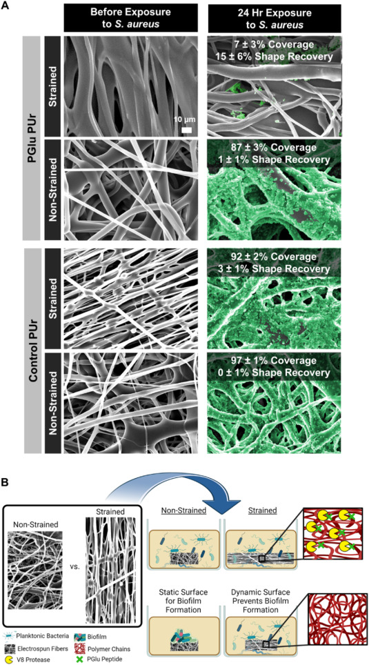
An interesting concept in preventing bacteria from forming a biofilm has been tested for a bacteria-responsive wound dressing. Gunderson et al (2024) electrospun a polyglutamic acid-containing polyurethane (PGlu PUr) membrane and induced strain in the membrane by depositing on a rotating collector. The polyglutamic acid (PGlu) incorporated into the polymer is susceptible to degradation by V8 protease released by Staph. aureus. Enzymatic cleavage of PGlu in the backbone by V8 caused the release of the strain in the membrane and the subsequent recovery of the membrane disrupted bacteria adhesion and inhibited biofilm formation. The same effect was seen with cultures of Staph. aureus, Staph. epidermidis, and E. coli on the membrane. On controlled, non-strained PGlu PUr membrane, no recovery of the membrane was seen as biofilm formed on its surface. The inhibition by the strained PGlu PUr membrane was observed in a 24 h incubation where bacteria coverage was only about 7% while non-strained membranes were completely covered with biofilm (87 - 97%).
(A) Biofilm formation on PGlu PUr and Control PUr fibers. Scanning electron micrographs of strained and unstrained fibers (left) before exposure to Staph. aureus and (right) after 24 ?h of exposure to Staph. aureus in biofilm forming conditions. Scale bar applies to all images. Quantified 2D biofilm coverage and shape recovery is overlaid on each image. Biofilms were artificially colored green using GIMP software. (B) Schematic representation of mechanism for biofilm disruption on strained PGlu PUr fibers [Gunderson et al 2024].
Bacteria trap
An interesting application of electrospun membrane is its potential ability to attract bacteria from the wound to the dressing. Abrigo et al (2015b) showed that surface modification of electrospun polystyrene with alkylamine was able to enhance attachment of E. coli. Conceptually, it may be able to draw bacteria from the wound and removed when the dressing is changed. However, more tests are needed to check the performance and viability of this strategy.
Hemostasis
In an accident or battlefield, it is vital that a wound dressing is able to control bleeding from the injury. A good hemostatic wound dressing goes beyond absorption of fluid, it would facilitate blood clotting to quickly arrest hemorrhaging. Saqr et al (2025) constructed electrospun polyacrylonitrile (PAN) nanofiber mats loaded with hemostatic agents, exfoliated bentonite (exben) and calcium chloride (CaCl2 for use as hemostatic material. The resultant electrospun PAN/exben/CaCl2 mat demonstrated a clotting time of just 105 s, a reduction of more than half the time taken for commercial cotton and gauze bandages which both took 270 s. The superior performance of the PAN/exben/CaCl2 mat can be attributed to the high surface area of water absorption ability of bentonite and its significant cation exchange capacity. The addition of CaCl2 facilitated enzyme catalysis during the hemostasis process through the supply of calcium ions and contributed to fibrin formation.
Application
Conventional application of wound dressing is to have the dressing packed in a sterile pouch. When there is a need to use the dressing, it is taken out of the pouch and applied directly onto the wound. Similarly, electrospun wound dressing may also be used in the same way.
Palo et al (2019) constructed a bi-layered wound dressing with a solvent cast (SC) bottom layer and an upper layer consisting of either electrospun fibers or three-dimensional (3D) printing. Both layers were made from a blend of polyvinyl alcohol (PVA) and sodium alginate (SA). Adhesion tests showed that cross-linked electrospun layer significantly reduces adhesiveness compared to 3D printed macroporous layer and SC film. This is advantageous in wound dressing as it allows easy removable from damaged tissues. Their study showed that SC/3D printed dressing adhesive behaviour is similar to SC thus the 3D printed layer offers no advantage in terms of adhesion. Tests on cell biocompatibility using fibroblasts also showed no significant difference in the cell viability between SC/3D printed dressing and SC film. However, with the electrospun layer, cell viability was greater. Therefore, having an electrospun layer on SC film was shown to offer several advantages for wound dressing.
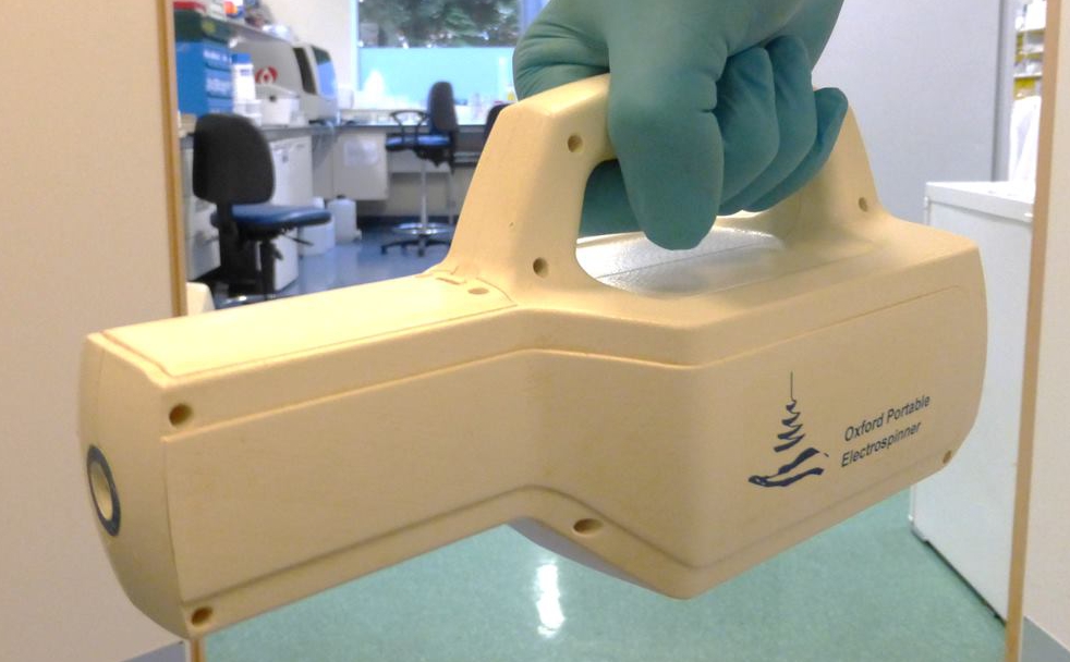
Portable electrospinning devices may offer an alternative way of covering the wound. The device typically runs on batteries to generate the required high voltage for initiating electrospinning from a cartridge filled with the solution. Mouthuy et al (2015) described the details of their portable electrospinning device which have been shown to be capable of producing nanofibers from a wide range of polymers such as polycaprolactone, polyvinyl alcohol, polyethylene oxide and poly(vinyl butyral) to name a few. Their battery powered device is able to run for 100 minutes at 13 kV. Feasibility of the device in depositing fibers on skin was demonstrated on human volunteers and pig skin [Mouthuy et al 2015].
Portable electrospinning device. Photo credit: P-A Mouthuy, University of Oxford, UK
In a demonstration of wound coverage using in situ precise gas assisted electrospinning, Lv et al (2016) covered a simulated open head injury with electrospun N-octyl-2-cyanoacrylate (NOCA), a commercial tissue adhesive (medical glue), fibers. The NOCA nanofibrous membrane covering the hole showed no fluid leakage. A wound area of 4-9 cm2 can be covered by a layer of nanofibrous membrane with 20s of electrospinning. The video below showed the wound coverage using electrospinning.
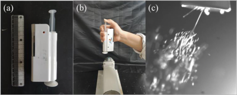
Liu et al tested the feasibility of using a hand-held portable electrospinning apparatus, HHE-1 from Qingdao Junada Technology Co., Ltd for deposition of fibers on a wound. This simple electrospinning apparatus ejects the solution through a syringe manually by the user's thumb. A disk covering the solution exit presumably to direct the electrospinning jet. Liu et al (2018) used polymers, poly(vinyl pyrrolidone) (PVP) and poly(vinyl butyral) (PVB) as the polymer carrier with iodine and iodine complex as anti-bacterial agents for electrospinning onto wound. PVP and PVB were both soluble in ethanol thus safe to use for direct application on wound. Comparison of the anti-bacterial properties of iodine-based electrospun membranes against E. coli and S. aureus showed that PVP with iodine had the best inhibition property and PVB/poly(vinylpyrrolidone)-iodine complex the least. The portable hand-held electrospinning device was able to deposit a layer of fibers on a hand and the fibers were sufficiently compact such that the deposited membrane may be peeled off from the hand.
The handheld electrospinning apparatus (a) and the in situ electrospinning process (b). The electrospinning jets can be seen from the spinneret [Liu et al 2018]
Using the same handheld device, HHE-1 from Qingdao Junada Technology Co., Ltd, Zhou et al (2023) loaded the electrospinning device with a polyvinylpyrrolidone (PVP) solution containing zein/Yunnan Baiyao (YB) particles and ciprofloxacin (CIP). CIP is a broad spectrum antibacterial medication while YB is a traditional Chinese herbal preparation for treating bruises, injuries, and bleeding wounds. When applied on a wound, fast dissolution of PVP (within a minute) would release CIP for disinfection. Since zein/YB particles are insoluble in water, they were released when the PVP dissolved. Slower release of YB from zein over 12 h would facilitate hemostatic and myogenic effects for wound healing. YB also showed antibacterial effect through slow release from zein with longer incubation time leading to greater antibacterial effect. Inhibition of Wb800 and Escherichia coli increases from 88.3% to 99.9% and from 81.1% to 99.9% after 2 and 12 h, respectively. Therefore, having both CIP and YB encapsulated in different forms has the synergistic effect of addressing short term and longer term healing of the wound.
In a demonstration of the versatility of a handheld electrospinning device, Xu et al (2022) was able to deposit polyvinyl alcohol (PVA) fibers containing bone marrow-derived stem cells (BMSCs) onto full-thickness skin wounds. The PVA solution was prepared using phosphate-buffered saline (PBS) and 1x10⁷ BMSCs were added to the solution prior to electrospinning. Cell viability test using dead/live staining showed 90.15% survival rate immediately after electrospinning. In vivo tests using SD rats demonstrated significantly faster wound healing in the PVA/cell group compared to PVA only and untreated control. On day 14, the wound covered with the PVA/cell group was almost completely closed. The epidermis was completely covered by epithelial tissue with normal skin appendages found around the wound. Healing was slower in the PVA only group with the subcutaneous tissues thinner than the PVA/cell group. The group without any intervention were still in the granulation tissue repair state with many capillaries, fibroblasts and inflammatory cells. Zhou et al (2024) used a portable electrospinning device to directly apply bilayered fiber scaffold on the skin with the outer layer composed of hydrophobic polycaprolactone (PCL) and a hydrophilic inner layer composed of gelatin, poly(L-lactic acid) (PLLA), and magnesium oxide nanoparticles (MgO NPs). At optimal concentration of MgO NPs, wound healing on Sprague Dawley (SD) rats was significantly better than scaffolds with lower and higher amounts of MgO NPs. This has been attributed to the antibacterial properties of MgO which inhibited colonization and growth of bacteria on the wound. Complete wound healing was observed on day 14. However, in vitro studies using human umbilical vein endothelial cells (HUVECs) and NIH/3T3s showed reduced viability at high concentration of MgO NPs. With excessive Mg ion concentration, in vivo studies showed a slower rate of healing compared to the control group at all time points.
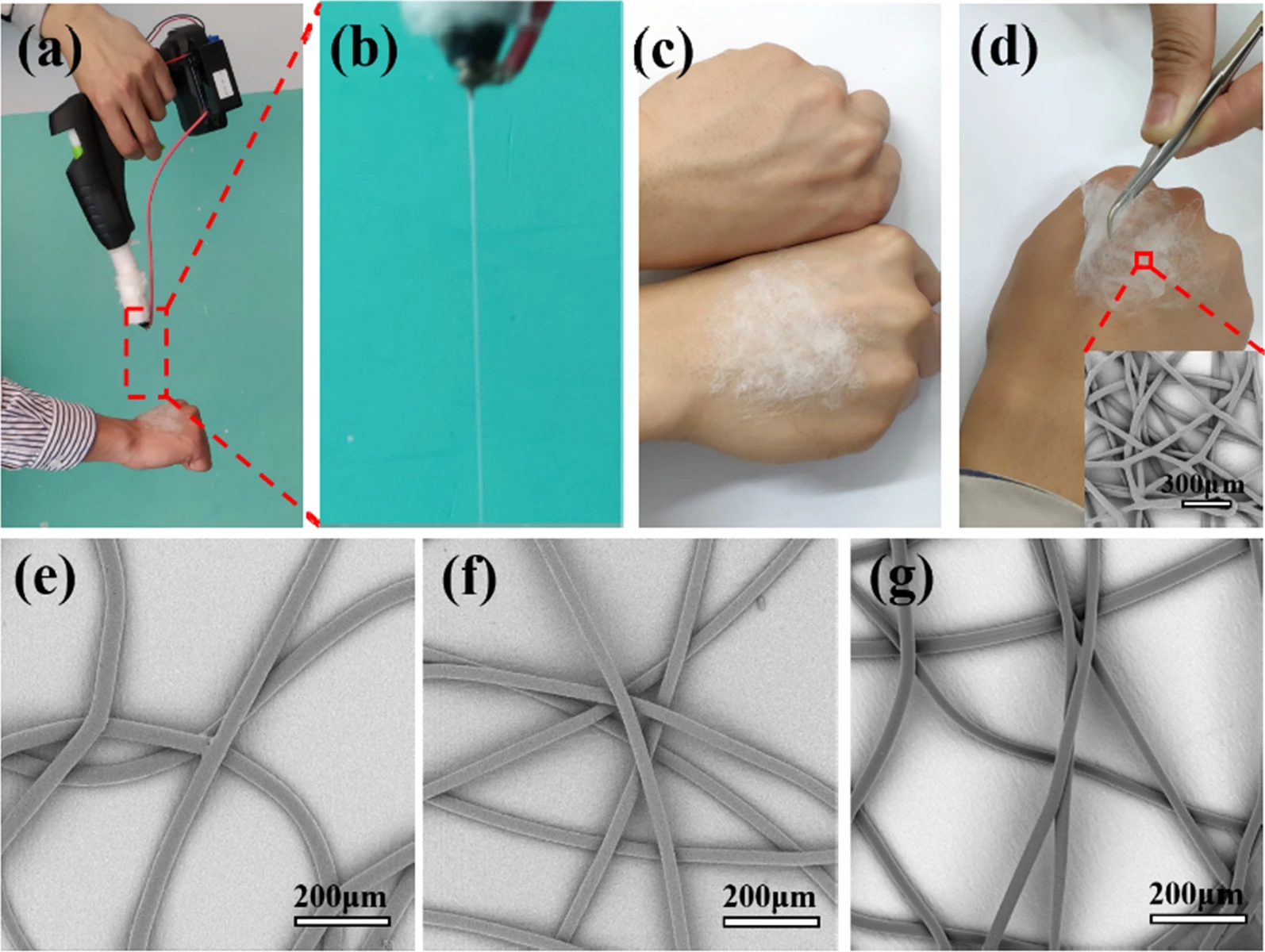
One main concern about direct electrospinning application onto wounds is the presence of solvents. If electrospinning is carried out by melt polymer, there will be no solvent and it can be safely applied onto the wound. For a portable melt electrospinning device, the challenge is to insulate the heating element from the high voltage. In a laboratory setup, it is possible to physically isolate the heating element from the high voltage. In a portable device, the space constraints means that both parts will be at close proximity to one another. A unique material which has excellent heat conductivity but electrically insulating property is the key to construct a portable melt electrospinning device. This allows the heat to be conducted close to the nozzle tip where the high voltage is applied while insulating the heating element from the high voltage source. Aluminum nitride (AlN) is one such material with good electrical insulation and heat transfer capacity. Zhao et al (2020) showed that with an AlN tube to conduct the heat within the setup, they were able to melt electrospin poly(lactic acid) (PLA), poly(lactic-co-glycolic acid) (PLGA), polycaprolactone (PCL), and hot-melt adhesive. The device was tested on a mouse for direct fiber deposition on a cut wound. The PCL fiber mesh that was melt electrospun on the wound was able to prevent blood from spilling out.
Lucian et al (2025) conducted a clinical study of SpincareTM, a portable electrospinning device for direct application of nanofibers matrix on wound surface. The nanofiber material is made of proprietary medical-grade polyurethane-based polymer and is designed to naturally detach as the wound heals. Comparison was made between patients with standard negative pressure wound therapy (NPWT) followed by conventional care (control group) and NPWT followed by SpincareTM applied nanofibers (study group). The clinical result showed that the SpincareTM group reported greater rate of complete pain relief by day 10 (46.6%) compared to the control group (6.6%) which can be attributed to protective, non-adherent properties of the nanopolymer film. The greater protection offered by the water-resistant properties of the film allow most patients to resume hygiene routines without compromising wound protection translated to lower rehospitalization rates for the Spincare group (23.3%) compared to the control group (60%). Hence the use of portable electrospinning devices such as SpincareTM may offer significant advantages in clinical applications.
Optical pictures showing the process of melt e-spinning PCL fibers directly onto the skin producing by the hand-held melt e-spinning apparatus in 5 min and SEM images of fibers with various polymer materials produced by the hand-held melt e-spinning apparatus to further test the performances of the apparatus. (a) The apparatus was operated by one hand and the other hand receives the PCL fibers. (b) Magnified view of spinning jet. (c) Comparison of two hands with or without fiber membrane. (d) The picture shows the e-spun fiber membrane has good flexibility and the inset SEM picture is the e-spun fibers. e PLA fibers. f PLGA fibers. g hot-melt adhesive fibers [Zhao et al 2020].
Multi-functionality
The versatility of electrospinning means that the resultant fibers can exhibit multiple functionality to aid wound healing. Core-shell fibers can be easily constructed such that the core and sheath material have different role to play in the wound dressing.
Zhan et al (2022) constructed multifunctional core-shell electrospun fibers for the purpose of diabetic wound healing. Four different materials were used in the fibers. Chitosan (CS) which exhibits antibacterial activity was used as the shell material so that the dressing would inhibit bacteria found on the wound. The core contains bioactive compounds, copper (Cu) salt and decellularized Wharton's jelly matrix (DWJM), and poly(L-Lactide-co-caprolactone) (PLCL). Cu is known to promote angiogenesis and endothelial migration and this will help wound healing at the intermediate stage. DWJM contains a natural extracellular matrix (ECM) and a source of endogenous growth factors to promote collagen deposition and wound healing. PLCL was added into the core matrix to provide stable electrospinning and mechanical strength for the composite fiber. In vivo test carried out using Sprague-Dawley (SD) rats diabetic model showed that the CTS/PLCL/DWJM/Cu core-shell nanofibers wound dressing exhibited the fastest healing compared to other wound dressings with one or more active substances absent. Skin appendages including hair follicles were evident at day 16 for the CTS/PLCL/DWJM/Cu core-shell nanofibers wound dressing group while healing for the other groups were slower.
Multiple spinnerets with each spinning a material with a particular functionality may be used to form a mat with a mixture of fibers. Du et al (2017) used a dual spinnerets electrospinning setup to form a membrane with antibacterial and wound healing properties. In their setup, electrospun fibers were made out of poly(vinyl alcohol) (PVA)/silver nanoparticles (AgNPs) and poly(caprolactone) (PCL)/ascorbyl palmitate (AP). The silver nanoparticles (AgNPs) are the antibacterial agent while AP aids wound healing. In vitro cytotoxicity test using 3T3 fibroblasts showed that proliferation were reduced with electrospun PVA/PCL/AgNPs compared to neat PVA/PCL. With PVA/PCL/AgNPs/AP, the fibroblast proliferation was 87% of the control which demonstrates the benefit of the presence of AP. In vivo studies using a rat model showed that wound closure from PVA/PCL/AgNPs/AP electrospun fibrous mat has the best result compared to all other groups including PVA/PCL/AgNPs, PVA/PCL/AP and PVA/PCL mesh. Li et al (2018) created a multi-functional wound dressing by creating a double layer electrospun membrane. The inner layer that comes into contact with skin was made of electrospun chitosan for its biocompatibility and and intrinsic antibacterial nature. Electrospun polycaprolactone (PCL) fibers were used as the outer layer to instill mechanical strength to the membrane. Anti-bacterial compounds, lidocaine hydrochloride (LID) and mupirocin was added to chitosan and PCL fibers respectively. The release profiles of LID and mupirocin were very different. 66% of LID was released in the first hours followed by gradual release to 85% in the following 6 hrs. Mupirocin showed an initial release of 57% in the first 6 hrs followed by sustained release of another 30% over the next 5 days. Therefore it can be seen that LID was released very quickly during the initial phase while mupirocin was able to maintain a gradual release for the remaining half of its load after the first 6 hrs. Slower release of mupirocin was attributed to stronger chemical linkage with PCL molecules. Such drug release profile is clinically relevant to achieve therapeutic concentration of the drug in minimal time.
Schulte-Werning et al (2021) used blending to incorporate several functionalities into the electrospun fibers. The ingredients forming the blend were chloramphenicol (CAM), an antibiotic, water-soluble β-1.3/1.6 glucan (SBG®), an active ingredient in the topical treatment of diabetic foot and leg ulcers, chitosan (CHI), a bioactive polymer which exhibits intrinsic antimicrobial activity, hydroxypropylmethylcellulose (HPMC), a cellulose-based polymer with high swelling capacity, and polyethylene oxide (PEO) to facilitate fiber formation from electrospinning. Instead of using conventional nozzle electrospinning, an Elmarco NanospiderTM NS Lab machine with electrode wire as the spinneret. The resultant electrospun multi-functional nanofibers were shown to inhibit E. coli and S. aureus, non-toxicity towards keratinocytes (HaCaT cells) and macrophages (RAW 264.7), and exhibit anti-inflammatory activity. Interestingly, in the sample that contains CHI but not CAM, no antimicrobial effect was seen and this has been attributed to the neutral pH of the gel. CHI requires an acidic environment for protonation so as to exhibit antimicrobial effect. Although CHI does not contribute to antimicrobial functionality in this wound dressing, its presence increases the mechanical strength and improves the anti-inflammatory of the membrane in the presence of SBG®. Upon exposure to fluid, this multi-functional nanofiber membrane showed a high swelling index and became transparent. This helps to adhere to the wound and maintain a moist local environment. High transparency of the membrane also allows examination of the wound without the need to remove the dressing.
A wound dressing may be constructed from different materials or processes to take advantage of the properties provided by each structure. Liu et al (2023) constructed a skin substitute using multi-layers of electrospun polycaprolactone (PCL) fibers and sprayed alginate hydrogel powder by alternating electrospinning and spraying. The multi-layered structure was wetted and cross-linked so that the alginate formed a stable matrix with the PCL fibrous layers to form a fiber hydrogel interpenetrated network (FHIPN). Amino-terminated hyperbranched polyamide (ATHBP) with antibacterial properties was added to the FHIPN by dipping the FHIPN in ATHBP solution to form functionalized fiber-hydrogel interpenetrating network (FFHIPN). The amino groups on ATHBP would bind to the carboxyl groups of sodium alginate through Coulomb interaction. In this setup, the PCL fibrous layers would provide the necessary elasticity and mechanical strength. On a wound injury, the alginate hydrogel would absorb the wound exudate and swell to create a more open FHIPN structure which encourages cell infiltration and at the same time provides a wet environment for wound healing. In vivo study using mouse full-thickness wound defect model showed significantly better wound healing using FHIPN and FFHIPN compared to the negative control using Gauze. In particular, histological tests showed the appearance of hair follicle structures on FHIPN and FFHIPN treated mice by day 7 while a small amount of hair follicle appeared in the wounds treated with Gauze on day 14. At day 14 there was almost complete wound closure for FFHIPN treated wounds.
Huang et al (2023) constructed an multilayered electrospun membrane with a Curcumin/Gelatin (Cur/Gel) as middle layer sandwiched between Gentamicin/Polyvinyl alcohol (Gt/PVA) as outer layers. Cur is a natural polyphenolic compound with anticancer, antioxidant, anti-inflammatory, and antibacterial properties and it was added to the electrospun wound dressing to promote healing. Gentamicin (Gt) is an antibiotic added to the membrane to prevent infection. PVA was used as a carrier for Gt and for absorbing wound exudate. To prevent the dissolution of PVA, the electrospun multilayered membrane was cross-linked using citric acid (CA). The wound dressing was found to be effective against Staphylococcus aureus and Escherichia coli although in the absence of Gt, Cur alone was not effective in inhibiting both bacteria. In vitro culture of L929 mouse fibroblasts showed no cytotoxicity effect. Interestingly, the adhesion of L929 cells on the membrane surface was poor with the cells maintaining a spherical morphology after 2 days. This may be an advantage as a wound dressing as the resistance to cell adhesion and may help prevent secondary damage during wound replacement.
In vivo
In vivo studies are critical to examine the efficacy of the device in the complex physiological environment. It is also an opportunity to test the actual application of the device. Shabunin et al (2019) tested the feasibility and efficacy of a bilayer electrospun dressing for treatment of burnt wound. The two electrospun layers comprised of a non-biodegradable alcohol-soluble aliphatic copolyamide (CoPA) (copolymer of poly(ε-caprolactam) [-NH-(CH2)5-CO-]n and poly(hexamethylenediamineadipinate) [-NH(CH2)6NHCO(CH2)4CO-]n) layer to provide mechanical support and a degradable chitosan/chitin layer to facilitate wound healing. The CoPA electrospun layer is separated when the dressing is removed leaving behind the chitin/chitosan layer to facilitate the epithelialization of the wound. In their In vivo study using third degree burn model on rats, almost complete (up to 97.8%) epithelialization of the wound surface had been achieved within 28 days for the group with the bilayer electrospun dressing. There were no deaths or purulent complications in the target group while there are 25% and 59.7% cases respectively in the control group. With electrospun CoPA membrane only, there were no deaths but 11% purulent complications. The bilayer electrospun wound dressing have shown the potential for treatment of burn wounds.
Another area of investigation is the type of active compounds added to the scaffold to promote wound healing. Bioactive compounds such as growth factors and others are often incorporated into electrospun wound dressings to accelerate the healing process. Losi et al (2020) compared the efficacy of wound healing using electrospun fibrin-based scaffold loaded with platelet lysate (PL). PL is derived from freeze-thawing cycles of platelet concentrates from peripheral blood and this contains a complex composition of growth factors and other biomolecules. Tests on a full thickness skin wound diabetic mice showed that fibrin-based scaffold loaded with PL performed better than electrospun scaffold loaded with growth factors, neat electrospun scaffold and Mepore® after 14 days. In the construction of the electrospun fibrin-based scaffold, fibrinogen solutions were first used to electrospun into fibers. Thrombin was subsequently sprayed onto the fibrinogen fibers and incubated for 30 min to allow fibrin polymerization. This method of preparing fibrin fibers allows bioactive compounds to be loaded as there are no post-electrospinning washing or chemical cross-linking steps.
Dhondale et al (2024) investigated the effect of multi-layered electrospun wound dressing loaded with heparin sodium (HS) and TPGS-1000 to promote healing of wounds in diabetes. The multi-layered wound dressing was made of a core layer of electrospun gelatin (gel) membrane loaded with HS and TPGS-1000 sandwiched between electrospun polycaprolactone (PCL)/gel membranes. The hydrophobicity of PCL provided a barrier to control the release of HS and TPGS-1000 by reducing the initial burst release and prolonging the release to 24h. When tested on a diabetic rats model, there was significant wound healing on the wound dressing applied group compared to blank nanofibers dressing and marketed cream treated groups on day 14 and 21. This demonstrated the benefits of having nanofibers wound dressings with controlled drug release in treatment of diabetic wounds.
There are numerous researches on the use of electrospun membranes for wound healing. The relative ease of loading electrospun membrane with drugs may also render it useful for the treatment of skin cancer. Song et al (2024) developed multi-functional bilayered nanofibers composite for postoperative wound healing of cutaneous squamous cell carcinoma. For the bilayered scaffold, the outer layer was made of electrospun hydrophobic polycaprolactone (PCL) membrane loaded with antibacterial enrofloxacin (ENR) while the inner layer was made of electrospun hydrophilic PCL-gelatin (PCL-Gel) nanofibers membrane loaded with anticancer drug bleomycin (PG-BLM). In vitro study showed initial burst release of the drugs which create an initial aseptic condition and antitumor effect followed by more gradual and sustained release after 24 h. The ENR loaded PCL membrane showed antibacterial effect on E. coli and S. aureus. Both BLM and ENR loaded membranes did not affect the adhesion or growth of normal keratinocyte (HaCaT) cells. Human epidermoid carcinoma cells (A431) cultured on PCL-ENR and PG scaffold did not show any changes in viability over 5 days but on PG-BLM, there were significant non-viable cells. On a partial tumor resection and skin defect models in immunodeficient mice, the PCL-ENR@PG-BLM group showed good wound healing and significantly lower expression level of Ki-67 in the skin tissue compared to PCL-PG group and the control group. The lower expression level of Ki-67 indicated inhibition of the growth of skin tumor cells and tumor metastasis.
Published date: 17 November 2015
Last updated: 26 May 2026
Abrigo M, Kingshott P, McArthur S L. Electrospun Polystyrene Fiber Diameter Influences Bacterial Attachment, Proliferation and Growth. ACS Appl Mater Interfaces 2015; 7: 7644.
Abrigo M, Kingshott P, McArthur S L. Bacterial response to different surface chemistries fabricated by plasma polymerization on electrospun nanofibers. Biointerphases 2015b; 10: 04A301.
Abrigo M, McArthur S L, Kingshott P. Electrospun Nanofibers as Dressings for Chronic Wound Care: Advances, Challenges, and Future Prospects. Macromol. Biosci. 2014; 14: 772.
Ahire J J, Neveling D P, Hattingh M, Dicks L M T. Ciprofloxacin-Eluting Nanofibers Inhibits Biofilm Formation by Pseudomonas aeruginosa and a Methicillin-Resistant Staphylococcus aureus. PLoS ONE 2015; 10: e0123648.
Open Access
Al Kayal T, Losi P, Pierozzi S, Soldani G. A New Method for Fibrin-Based Electrospun/Sprayed Scaffold Fabrication. Sci Rep 2020; 10: 511.
Bao X, Zhu Q, Chen Y, Tang H, Deng W, Guo H, Zeng L. Antibacterial and antioxidant films based on HA/Gr/TA fabricated using electrospinning for wound healing. International Journal of Pharmaceutics 2022; 626: 122139.
Open Access
Brooker C, Richard D'Arcy R Mele E, Willcock H. Designing responsive dressings for inflammatory skin disorders; encapsulating antioxidant nanoparticles into biocompatible electrospun fibres. Soft Matter 2021; 17: 3775.
Open Access
Cai Z X, Mo X M, Zhang K H, Fan L P, Yin A L, He C L, Wang H S. Fabrication of Chitosan/Silk Fibroin Composite Nanofibers for Wound-dressing Applications. Int. J. Mol. Sci. 2010; 11: 3529.
Open Access
Chaudhary A, Gupta A, Mathur R B, Dhakate S R. Effective antimicrobial filter from electrospun polyacrylonitrile-silver composite nanofibers membrane for conducive environment. Adv. Mat. Lett. 2014; 5: 562.
Chellamani K P, Balaji R S V, Veerasubramanian D. Development of Wound Dressing Made of Electrospun Tetracycline Hydrochloride Drug Incorporated PCL (Poly (ε-Caprolactone)) Nanomembrane. International Journal of Emerging Technology and Advanced Engineering 2014; 4: 251.
Open Access
Dhondale M R, Manjit M, Jha A, Kumar M, Bharti K, Kumar D, Mishra B. Heparin sodium enriched gelatin/polycaprolactone based multi-layer nanofibrous scaffold for accelerated wound healing in diabetes. RSC Pharm. 2024; 1: 1021.
https://pubs.rsc.org/en/content/articlehtml/2024/pm/d4pm00130c Open Access
Du L, Xu H Z, Li T, Zhang Y, Zou F Y. Fabrication of ascorbyl palmitate loaded poly(caprolactone)/silver nanoparticle embedded poly(vinyl alcohol) hybrid nanofibre mats as active wound dressings via dual-spinneret electrospinning. RSC Adv. 2017; 7: 31310-31318.
Open Access
Gürtler A L, Sirois J P, Lang J C, Melican K, Rades T, Heinz A. Electros pun dressings with a dual release functionality of two anti-inflammatory active ingredients. RSC Pharm. 2024; 1: 570.
https://pubs.rsc.org/en/content/articlehtml/2024/pm/d4pm00147h Open Access
Hassiba AJ, El Zowalaty ME, Webster TJ, Abdullah AM, Nasrallah GK, Khalil KA, Luyt AS, Elzatahry AA. Synthesis, characterization, and antimicrobial properties of novel double layer nanocomposite electrospun fibers for wound dressing applications. International Journal of Nanomedicine 2017:12; 2205.
Open Access
Jones V, Grey J E, Harding K G. Wound dressings. BMJ 2006; 332: 777.
Open Access
Kehail A A and Brigham C J J. Anti-biofilm Activity of Solvent-Cast and Electrospun Polyhydroxyalkanoate Membranes Treated with Lysozyme. Polym Environ 2017 Article in press
Leonarta F and Lee C K. Nanofibrous Membrane with Encapsulated Glucose Oxidase for Self-Sustained Antimicrobial Applications. Membranes (Basel). 2021; 11(12): 997.
Open Access
Lev J, Holba M, Kalhotka L, Mikula P, Kimmer D. Improvements in the Structure of Electrospun Polyurethane Nanofibrous Materials Used for Bacterial Removal from Wastewater. International Journal of Theoretical and Applied Nanotechnology 2012; 1: 16.
Open Access
Li X, Wang C, Yang S, Liu P, Zhang B. Electrospun PCL/mupirocin and chitosan/lidocaine hydrochloride multifunctional double layer nanofibrous scaffolds for wound dressing applications. International Journal of Nanomedicine 2018; 2018: 5287.
Open Access
Liu G S, Yan X, Yan F F, Chen F X, Hao L Y, Chen S J, Lou T, Ning T, Long Y Z. In Situ Electrospinning Iodine-Based Fibrous Meshes for Antibacterial Wound Dressing. Nanoscale Res. Lett. 2018: 13: 309.
Open Access
Liu X, Gao L, Fu S, Zhao W, Wang F, Gao J, Li C, Wu H, Wang L. Polycaprolactone nanofiber-alginate hydrogel interpenetrated skin substitute for regulation of wound-substitute interface. Materials & Design 2023; 227: 111706.
Open Access
Liu Y, Franco A, Huang L, Clark R, Rafailovich M. Directing Cell Migration by Electrospun Fibers. NSTI-Nanotech 2010; 1: 881.
Losi P, Al Kayal T, Buscemi M, Foffa I, Cavallo A, Soldani G. Bilayered Fibrin-Based Electrospun-Sprayed Scaffold Loaded with Platelet Lysate Enhances Wound Healing in a Diabetic Mouse Model. Nanomaterials. 2020; 10(11):2128.
Open Access
Lucian BI, Cheregi CD, Sebastian HM, Ruxandra-Florina B, Maghiar L, Ilarie B, Anca H, Sachelarie L, Mircea-Ioan S. Electrospun Nanofibers in Wound Healing: Real-World Evaluation of SpincareTMTechnology. Bioengineering. 2025; 12(5):500.
https://www.mdpi.com/2306-5354/12/5/500 Open Access
Lv F Y, Dong R H, Li Z J, Qin C C, Yan X, He X X, Zhou Y, Yan S Y, Long Y Z. In situ precise electrospinning of medical glue fibers as nonsuture dural repair with high sealing capability and flexibility. International Journal of Nanomedicine 2016; 11: 4213. Open Access
Ma Z, Ji H, Tan D, Teng Y, Dong G, Zhou J, Qiu J, Zhang M. Silver nanoparticles decorated, flexible SiO2 nanofibers with long-term antibacterial effect as reusable wound cover. Colloids and Surfaces A: Physiochem. Eng. Aspects 2011; 387: 57.
Motealleh B, Zahedi P, Rezaeian I, Moghimi M, Abdolghaffari A H, Zarandi M A. Morphology, drug release, antibacterial, cell proliferation, and histology studies of chamomile-loaded wound dressing mats based on electrospun nanofibrous poly(?-caprolactone)/polystyrene blends. J. Biomed Mater. Res B Appl. Biomater. 2014; 102: 977.
Mouthuy P A, Groszkowski L, Ye H. Performances of a portable electrospinning apparatus. Biotechnology Letters 2015; 37: 1107.
Open Access
Palo M, Rönkönharju S, Tiirik K, Viidik L, Sandler N, Kogermann K. Bi-Layered Polymer Carriers with Surface Modification by Electrospinning for Potential Wound Care Applications. Pharmaceutics 2019; 11(12): 678.
Open Access
Queen D, Gaylor J D S, Evans J H, Courtney J M. The preclinical evaluation of the water vapour transmission rate through burn wound dressings. Biomaterials 1987; 8: 367.
Saqr B, Alqassar Bani Almarjeh R, Atassi Y. Development of an innovative cost-effective hemostatic material based on electrospun polyacrylonitrile/exfoliated bentonite/calcium chloride nanocomposite. Sci Rep 2025; 15: 15795.
https://www.nature.com/articles/s41598-025-00569-3 Open Access.
Schulte-Werning LV, Murugaiah A, Singh B, Johannessen M, Engstad RE, Skalko-Basnet N, Holsæter AM. Multifunctional Nanofibrous Dressing with Antimicrobial and Anti-Inflammatory Properties Prepared by Needle-Free Electrospinning. Pharmaceutics. 2021; 13(9):1527.
Open Access
Shabunin A S, Yudin V E, Dobrovolskaya I P, Zinovyev E V, Zubov V, Ivan'kova E M, Morganti P. Composite Wound Dressing Based on Chitin/Chitosan Nanofibers: Processing and Biomedical Applications. Cosmetics 2019, 6(1), 16.
Open Access
Shang S, Yang F, Cheng X, Walboomers X F, Jansen J A. The Effect of Electrospun Fibre Alignment on the Behaviour of Rat Periodontal Ligament Cells. European Cells and Materials 2010; 19: 180.
Shin D, Kim M S, Yang C E, Lee W J, Roh T S, Baek W. Radially patterned polycaprolactone nanofibers as an active wound dressing agent. Arch Plast Surg. 2019; 46(5): 399.
Open Access
Song Y, Hu Q, Liu S, Yao G, Zhang H. Electrospinning drug-loaded polycaprolactone/polycaprolactone-gelatin multi-functional bilayer nanofibers composite scaffold for postoperative wound healing of cutaneous squamous cell carcinoma. Biomedical Technology 2024; 8: 65.
https://www.sciencedirect.com/science/article/pii/S2949723X2400031X Open Access
Unnithan A R, Barakat N A M, Pichiah P B T, Gnanasekaran G, Nirmala R, Cha Y S, Jung C H, El-Newehy M, Kim H Y. Wound-dressing materials with antibacterial activity from electrospun polyurethane-dextran nanofiber mats containing ciprofloxacin HCl. Carbohydrate Polymers 2012; 90: 1786.
Wang A, Xu C, Zhang C, Gan Y, Wang B. Experimental Investigation of the Properties of Electrospun Nanofibers for Potential Medical Application. Journal of Nanomaterials 2015; 2015: 418932.
Open Access
Xu S, Lu T, Yang L, Luo S, Wang Z, Ye C. In situ cell electrospun using a portable handheld electrospinning apparatus for the repair of wound healing in rats. International Wound Journal 2022 Early view.
Open Access
Yue Y, Liu X, Pang L, Liu Y, Lin Y, Xiang T, Li J, Liao S, Jiang Y. Astragalus Polysaccharides/PVA Nanofiber Membranes Containing Astragaloside IV-Loaded Liposomes and Their Potential Use for Wound Healing. Evidence-Based Complementary and Alternative Medicine 2022; 2022: 9716271.
Open Access
Zhan A, Chen L, Sun W, Tang Y, Chen J, Yu D, Zhang W. Enhancement of diabetic wound healing using a core-shell nanofiber platform with sequential antibacterial, angiogenic, and collagen deposition activities. Materials & Design 2022; 218: 110660.
Open Access
Zhao J, Han F, Zhang W, Yang Y, You D, Li L. Toward improved wound dressings: effects of polydopamine-decorated poly(lactic-co-glycolic acid) electrospinning incorporating basic fibroblast growth factor and ponericin G1. RSC Adv. 2019; 9: 33038.
Open Access
Zhao Y T, Zhang J, Gao Y, Liu X F, Liu J J, Wang X X, Xiang H F, Long Y Zl. Self-powered portable melt electrospinning for in situ wound dressing. J Nanobiotechnol 2020; 18: 111.
Open Access
Zhou J, Wang L, Gong W, Wang B, Yu D-G, Zhu Y. Integrating Chinese Herbs and Western Medicine for New Wound Dressings through Handheld Electrospinning. Biomedicines. 2023; 11(8):2146.
Open Access
Zhou T, Chen Y, Fu L, Wang S, Ding H, Bai Q, Guan J, Mao Y. In situ MgO nanoparticle-doped Janus electrospun dressing against bacterial invasion and immune imbalance for irregular wound healing. Regenerative Biomaterials 2024; 11: rbae107.
https://academic.oup.com/rb/article/doi/10.1093/rb/rbae107/7739756 Open Access.